 |
|
|
An e-mail conversation with Graeme Laver
|
 |
Graeme Laver (1929-2008) was Professor of Biochemistry and Molecular Biology at the Australian National University in Canberra. He and influenza virus have had a long intertwined history and he shared some of the story with us here.
Graeme died in 2008. After his death his friend and colleague Rob Webster wrote:
"Graeme Laver, the maverick of influenza research in Australia, was always prepared to challenge authorities. He established the biochemical basis of antigenic drift and shift in seasonal and pandemic influenza viruses and played a key role in the development of the anti-influenza drug Relenza. It was Graeme's contention that antiviral drugs (Relenza and Tamiflu) should be available in everyone's medicine cabinet. His argument is that many will die in an influenza pandemic before available stockpiles could be distributed.
He also contended that those claiming that this would promote antiviral resistance have got it wrong!
In classical Graeme style, he finished his life with a great flourish while on his way to a scientific meeting on influenza in Portugal. The air traffic controllers cleared the air space over Heathrow so that he could receive rapid medical attention. We his friends all know that he would have reveled in the mayhem caused had he been aware of it."
Those brief comments only give a glimpse of Graeme's character and his e-mail conversations with me should also indicate what fun it was to know him. He will be missed.
Some of the first questions I asked Graeme were what were the early influences on him and, in particular, how did he get started working on influenza and on neuraminidase.
He answered:
Alfred Gottschalk was working on RDE - the receptor destroying enzyme - from influenza virus at the Walter and Eliza Hall Institute in Melbourne. In about 1947 I was working as a technician at the same Institute while I did a part-time Science course at Melbourne University and spent some time working for Gottschalk , who though a naturalized Australian, had a very thick German accent and was called "Uncle Alfie". Gottschalk insisted, that if RDE were destroying receptors for flu virus on red cells there had to be a "split product" and he worked with obsessive zeal to find it. I was able to help him with experiments in which he digested ovomucin with RDE, dialysed the "split product" and eventually characterized it as sialic acid (N-acetyl neuraminic acid). In this way RDE became known as sialidase (or neuraminidase).
I completed a Ph.D. in London in 1958 and returned to Australia by the overland route to Bombay driving a small Standard 10 sedan. In the Bombay General Post Office I found a letter waiting for me offering me a job in the John Curtin School at the Australian National University. Here I teamed up with Stephen Fazekas de St Groth and his graduate student, Rob Webster, to work on the molecular structure and antigenic properties of the influenza virus. Very little was known about these things at this time. In 1958 the accepted method for disrupting flu virus was to shake the virus particles with ether. It quickly became apparent that ether did very little in the way of disrupting the virus and we found that the detergents, sodium deoxycholate and sodium dodecyl sulphate (SDS) did a much better job. This finding, in fact, formed the basis for many of the subsequent discoveries.
I asked Graeme why he chose to solve the structure of neuraminidase. He skipped over many years of work to answer:
In March 1977, I had organized a small meeting in Baden-near-Vienna on the Influenza Virus Hemagglutinin and traveled back to Canberra on the plane with Gillian Air. I said to her: "We can't have Skehel and Wiley have it all to themselves, when I get back I am going to crystallize the neuraminidase". Well, I did just that, but it was sheer luck and not at all intentional. I was, in fact trying to show that the Asian (H2N2) and the Hong Kong (H3N2) neuraminidases had similar sequences because Ed Kilbourne had claimed that antigenic similarity did not necessarily mean sequence similarity.
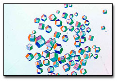
The N2 neuraminidase crystallized as thousands of small square plates when I dialysed the enzyme from a sucrose gradient against water. I told Gillian I had neuraminidase crystals. She exploded in scornful rage. "You stupid man", she said. "I've seen people like you before. All you have are salt crystals." I smiled. If either NaCl or sucrose had crystallized from distilled water I could re-write the chemistry books.
The crystals were neuraminidase alright. I didn't know what to do with them. I thought I might send them to Don Wiley in case he wanted to do the structure. Then an Australian immunologist, Alan Williams, who was working in Oxford, stopped by the lab, saw the crystals and said: "Those crystals must not go out of Australia", and introduced me to Carolyn Wright, a crystallographer at Sydney University. Carolyn collected the preliminary X-ray diffraction data on the neuraminidase crystals and then one evening we called her colleague Peter Colman who was in Munich and asked him if he would like to solve the structure. Peter came back to Melbourne to do this and the crystal structure of N2 neuraminidase was published in Nature in 1983 (J.N. Varghese, W.G.Laver and P.M.Colman Nature303:35-40, 1983).
 So there
you are! There was never any "choosing" to solve the NA structure. What started
as a scheme to be one up on Skehel and Wiley just sort of developed all by itself.
There are so many "what ifs... What if I had dialysed the enzyme against saline
instead of water? What if Alan Williams hadn't stopped in the lab that day?
What if Ed Kilbourne hadn't provoked me into looking at the sequences of Asian
and Hong Kong neuraminidases? I could go on and on.
So there
you are! There was never any "choosing" to solve the NA structure. What started
as a scheme to be one up on Skehel and Wiley just sort of developed all by itself.
There are so many "what ifs... What if I had dialysed the enzyme against saline
instead of water? What if Alan Williams hadn't stopped in the lab that day?
What if Ed Kilbourne hadn't provoked me into looking at the sequences of Asian
and Hong Kong neuraminidases? I could go on and on.
I wanted Graeme to place the work on neuraminidase in the context of the time. I asked what else was going on and how other discoveries affected his work.
He continued:
What was going on was the development of the influenza subunit vaccine. The flu vaccines in use in the 1950s contained virus particles inactivated with some agent, such as formaldehyde. These vaccines often produced toxic reactions when injected, sometimes described as worse than the disease itself. The toxicity was associated with the intact virus particle and people in Tommy Francis' lab in Ann Arbor found that ether-disrupted virus was much less toxic. This led to the proposition that the standard flu vaccine should be replaced by an ether-treated vaccine. When we found that sodium deoxycholate efficiently disrupted particles of influenza virus without affecting the hemagglutinin or neuraminidase activities, it was obvious that virus disrupted with this detergent might provide an effective non-toxic flu vaccine. This did indeed prove to be the case and the first commercial influenza "subunit" vaccine was made by the Australian Commonwealth Serum Laboratories in Melbourne. I believe that most, if not all, flu vaccines made today are so-called "subunit" vaccines.
We then discovered a quite amazing thing. Viruses disrupted with the detergent sodium dodecyl sulphate (SDS) at room temperature often retained hemagglutinin activity, others retained neuraminidase activity and some retained both. When these viruses were subjected to electrophoresis on cellulose acetate strips at room temperature in buffers containing about 1% SDS in one case pure hemagglutinin molecules and in the other case pure neuraminidase molecules migrated on the strips in the opposite way to all of the other virus proteins and could be eluted from the strips (if both HA and NA activities survived in SDS, then a mixture of HA and NA was obtained). I took preparations of hemagglutinin and neuraminidase made in this way to the National Institute for Medical Research at Mill Hill in London in 1969 where Robin Valentine examined them in the electron microscope both in the presence of SDS and after the removal of this detergent (Virology 38: 105-119, 1969). This showed very clearly that the hemagglutinin was a triangular rod-shaped molecule while the neuraminidase was mushroom-shaped with a head attached to long thin stalk with a small knob at the end. The head was square and box-shaped as Nick Wrigley so elegantly showed later but the description of the neuraminidase as "mushroom-shaped" may have been unfortunate. I saw recently a German film in which the neuraminidase was depicted as a molecule with a round plate-like head and stalk. When I told the producer the neuraminidase had a box-like head he retorted "You said it was mushroom-shaped, and I have never seen a square mushroom!"
This technique we discovered, of how to isolate pure, intact hemagglutinin and neuraminidase "spikes" from the virus, led to a number of other discoveries. John Skehel and Geoffrey Schild used it in the first demonstration that flu hemagglutinin consists of two polypeptides, HA1 and HA2 something which we also found at about the same time as well as showing that HA1 and HA2 were disulphide linked. Another key discovery was made in Ed Kilbourne's lab in New York where we showed that, following the mixed infection of cells with two different influenza A viruses, re-assortant viruses could be isolated which had the hemagglutinin from one parent and the neuraminidase from the other (Virology 30: 493-501,1966). This, in turn, led to Rob Webster's famous paper: "Antigenic Hybrids of Influenza A viruses with Surface Antigens to Order" (Virology 42: 633-642, 1970). These hybrid viruses were invaluable for much of the work which followed.
Graeme then gave a very brief description of why we think that neuraminidase activity is essential for flu virus.
What function did the neuraminidase play in the life cycle of the virus? In 1966, Joe Seto and Rudi Rott showed that the function of the neuraminidase was probably associated with the release of virus from host cells (Virology 30: 731-737, 1966). The following year, we also found that antibody directed specifically against flu neuraminidase did not prevent the infection of susceptible cells but prevented the release of newly formed virus particles. In 1974 this function of the enzyme was demonstrated most elegantly by Peter Palese, Dick Compans and their colleagues who showed electron micrographs of sections of cells infected with wild type virus and with a mutant which lacked neuraminidase activity (Virology 61:397-410,1974). In the latter case, the virus particles which budded off from the cell formed great aggregates still attached to the cell surface. It was clear then that without neuraminidase to release it, the virus was not going to go anywhere and the infection was effectively terminated. This finding was, of course, very encouraging when we started thinking about developing neuraminidase-inhibitors as a way to control flu.
Returning to 1966, Seto, Drzeniek and Rott showed that pronase treatment released a small molecular weight sialidase from H2N2 viruses. Would this sialidase be suitable for structural studies or was it all chewed up by the pronase? At first I was scornful ("You might just as well serve up a good steak after the dog has been at it !") but I was wrong and pronase-released neuraminidase provided the first crystals which opened the way for the rational design of potent and specific inhibitors of the enzyme.
And in answer to the general question of how structure led to biological insights Graeme went on to write:
Although the amino acid sequences of a number of flu A as well as flu B neuraminidases often differed greatly, Peter Colman and his colleagues noticed that some residues at various widely-separated positions along the linear neuraminidase polypeptide were totally conserved among all of the neuraminidase sequences of Type A and Type B influenza. Furthermore, when the linear polypeptide folded into the 3-dimensional structure of the active neuraminidase, these conserved residues all clustered together lining the walls of a deep cleft or canyon on the top surface of the neuraminidase. X-ray crystallography of neuraminidase crystals soaked in sialic acid showed sialic acid binding in this cleft which was clearly identified as the catalytic site. This site was thus shown to be totally conserved among all influenza viruses, indicating that if a substance could be found which specifically inhibited one type of influenza virus neuraminidase, then it would be effective against neuraminidases from all influenza virus strains, including those which have not yet appeared in man.
Some thought was given to making use of this conserved site to develop a universal influenza virus vaccine. However, that this approach would not work became evident when we determined the structure of epitopes on the neuraminidase. Peter Colman initiated this project. At the time, the structure of epitopes on proteins and the structures of the corresponding binding sites on antibodies were not known. Most assumed that epitopes on proteins contained 6 amino acid residues, but no direct evidence for this had been obtained.
We therefore set out to grow crystals of flu neuraminidase complexed with Fab fragments of monoclonal antibodies to the neuraminidase in order to determine, using X-ray crystallography, the 3-D structure of the complex. If successful. this would give the structure of the epitope on the neuraminidase and of the corresponding paratope on the antibody. This had not been done for any protein, but we knew that Roberto Poljack in Paris was trying to do the same thing, using hen egg lysozyme, and we were out to beat him. It was a real team effort. Rob Webster made the monoclonal antibodies and selected escape mutants of the neuraminidase, I produced the flu neuraminidase, made Fab fragments of Rob's antibodies, mixed the two and crystallized the resulting complexes. Peter Colman and his colleagues determined the structure of the complexes by X-ray crystallography and Gillian Air did much of the amino acid sequencing.
Peter, in fact, probably produced the first ever crystalline protein-antibody complex, even before the Paris crowd had one, but it is a sad story. Rob had made a small amount of monoclonal antibody to N2 neuraminidase called S10/1, and Peter grew crystals out of a mixture of N2 neuraminidase and S10/1 Fab. He sent me a photo of the thin crystals which were not big enough for X-rays and so we never knew if these were complex crystals or not. We never knew this because Rob Webster then went on holidays to some Canadian Lake for a couple of weeks and while he was away the freezer containing his S10/1 hybridoma cells broke down, and the cells were lost for ever and ever. Meanwhile Poljack grew crystals of lysozyme complexed with Fab and got the structure of this complex before we eventually got our neuraminidase-Fab structure (Colman, PM, Laver, WG, Varghese, JN, Baker, AT, Tulloch, PA, Air, GM and Webster, RG Nature 326: 358-363, 1987).
This showed that the epitope on the neuraminidase comprised 5 separate peptide segments with about 17 amino acid residues in contact with a similar number of amino acids in the antibody binding site. Single amino acid sequence changes in the peptide segments of the neuraminidase epitope rendered the neuraminidase invisible to the antibody so totally abolishing binding. This finding meant that even if antibody could be raised to that region on the neuraminidase which involved the conserved catalytic site, this antibody would still be susceptible to changes in the variable amino acids surrounding the conserved site and such a vaccine would not be effective against all strains of the virus. Small molecule inhibitors of the enzyme were therefore sought.
Attempts by others to identify such inhibitors by random screening failed. One drug company, for example, screened 25,000 compounds without coming up with a single inhibitor. Sialic acid, the substrate for neuraminidase, is itself a mild inhibitor of the enzyme, but the dehydrated compound, DANA (Neu5Ac2en), is a very much better inhibitor and in the 1970s, Peter Palese and his colleagues showed that DANA and some of its derivatives inhibited influenza virus replication in tissue culture but when tested in animals these compounds failed to prevent disease. Then, in the 1980s, Mark von Itzstein and his colleagues soaked sialic acid into crystals of flu neuramindase and determined the X-ray structure of the complex. This showed that opposite the 4-hydroxyl on the sialic acid there was a pocket in the neuraminidase, at the bottom of which were two glutamic acid residues. These glutamics were too far away from the substrate to play any role in catalysis but were nevertheless totally conserved among all flu strains. When the hydroxyl at the 4 position on DANA was replaced by an amino group the resulting compound was a better inhibitor than DANA but when 4-guanidino DANA was prepared and tested it was found to be a 1000-fold better inhibitor than DANA and this inhibition was specific for influenza neuraminidase and not for neuraminidases from other sources.
4-guanidino DANA is now known as Relenza. It has been successful in treating influenza in clinical trials and is now approved for use in Australia, the USA, Canada and in a number of other countries. Relenza, however, is not orally bioavailable. The very group which makes it such a good inhibitor, the guanidino group, also prevents Relenza from easily crossing membranes. It has to be administered as a powder which is inhaled into the lungs.
One of Graeme's best stories is not about structure, but it is an important part of the story of influenza - and well-worth reading!
The scene now shifts a little, from the lab into the field. In the 1960s, during my visits to the NIMR at Mill Hill, I became good friends with Helio Pereira who was Head of Virology and the WHO Influenza Centre. Helio was very interested in animal and avian influenza and we used to talk quite a bit about these viruses which until then had been isolated only from domestic animals or birds - apart from one incident. (In 1961 an H5N1 virus killed many terns in South Africa). Helio kept suggesting we go and look for flu in wild birds and of course the Great Barrier Reef was suggested as the ideal place to do flu research. (Can you think of somewhere better?)
 I went skiing
one winter with Helio at Chamonix and on the way there we called into WHO in
Geneva to talk to Martin Kaplan. Martin also was keen to look for flu in wild
birds and offered $500 towards the cost of a field trip if ever I needed it.
This must have been in January or February of 1969. So at the end of 1969 I
arranged a trip to Tryon Island. This is a deserted coral cay, about 50 miles
off the Queensland coast in the Capricorn Bunker group and in December is alive
with vast numbers of nesting wedge-tailed shearwaters and noddy terns.
I went skiing
one winter with Helio at Chamonix and on the way there we called into WHO in
Geneva to talk to Martin Kaplan. Martin also was keen to look for flu in wild
birds and offered $500 towards the cost of a field trip if ever I needed it.
This must have been in January or February of 1969. So at the end of 1969 I
arranged a trip to Tryon Island. This is a deserted coral cay, about 50 miles
off the Queensland coast in the Capricorn Bunker group and in December is alive
with vast numbers of nesting wedge-tailed shearwaters and noddy terns.
When I asked the head of my department for funds to look for flu in these birds, his reaction was "Laver is hallucinating". So the money from Martin came in very handy. The other comment from my departmental head was "Anyway, there is no way he is going to be able to catch the birds". Poor sod, I wasn't that stupid. I knew the shearwaters nested in burrows in the ground and all we had to do was bend over and pick them up. Now, I must admit the thought of those beautiful, healthy birds on a deserted coral island, surrounded by the bluest of blue seas under a scorching sun carrying influenza viruses was almost too bizarre to even contemplate seriously. But we were there, camped on the island for 3 weeks with all our food and water with us and no contact with civilization during the whole period. We set about looking for flu!
Instead of looking for virus we first looked for antibodies and set up gel-diffusion tests on the island in which we tested 201 sera from the shearwaters against flu A ribonucleoprotein. To my astonishment some of the sera gave precipitin lines! The precipitates were a bit diffuse and most of the lines were hard to see and I could never have published "Antibodies to flu A in shearwaters on Tryon Island" on the gel-diffusion tests alone. But they were enough to encourage us to do more tests back in the lab.
Now, most people when they looked for antibodies to flu in sera used the hemagglutination-inhibition tests. This test often gives false positives because of non-specific inhibitors and I chose to do neuraminidase-inhibition tests instead. The next question was: what neuraminidase to use in the tests? This was almost like trying to pick the winner in the Melbourne cup! We knew that Asian influenza N2 neuraminidase was present in a number of turkey viruses and we settled on this subtype for the tests. The test we used gave a bright red color if the enzyme was active and I will never forget the excitement we felt when one of the sera eliminated all the color!
We showed very quickly that this inhibition was due to antibody specific for N2 neuraminidase and we subsequently found, out of 320 shearwater sera tested, 18 which had N2 antibody. (Dasen and Laver , Bull. World Health Org 42, 885-889, 1970). This meant that these birds had in the past been infected with Type A flu and we set up more expeditions to try to isolate virus. The first virus was isolated from a tracheal swab taken from a wedge-tailed shearwater on Tryon Island in 1972 and in subsequent trips other viruses were isolated from shearwaters and white capped Noddy terns on Tryon Island and close-by Northwest Island. These birds were all completely healthy, even though the titres of virus in the swabs were quite high.
One of these viruses turned out to be of great importance. The 70th cloacal swab collected by one of the members of the expedition, Adrian Gibbs, yielded a virus which had a neuraminidase of a previously unknown subtype, N9, and this N9 neuraminidase gave the best crystals of any flu A or B neuraminidase so far examined. N9 neuraminidase crystals have now been used (not always successfully) by Gilead Sciences, Hoffman La Roche, BioCryst Pharmaceuticals, GlaxoWellcome, Eli Lilly, Abbott Labs., ZymeTx Corporation and Pfizer Ltd. in the design of novel neuraminidase inhibitors which these companies hoped to market as anti-influenza drugs.
Anyone who has wondered "why do I have to be vaccinated against influenza virus once again?" has an interest in the ability of this virus to undergo antigenic changes. These changes are called antigenic drift and antigenic shift. It is changes in the hemagglutinin that allow the virus to escape the previous year's needle stick. Graeme's involvement with influenza virus also included some of the early research on hemagglutinin - and he added the following to the history of drift and shift:
 When we (Rob
Webster, Stephen Fazekas and I) started working on flu in 1959 or so virtually
nothing was known about the structural proteins in the influenza virus particle.Polyacrylamide
gel electrophoresis had not been invented then and there was not even any information
how many proteins existed in the virus particle. We tried to get a handle on this
by looking for free N-terminal amino acids in purified preparations of flu virus.
We found two: aspartic acid and glycine. We then took pure HA which we isolated
by electrophoresis of virus particles disrupted with SDS and showed that this
was the protein which had the N-terminal aspartic and glycine . So this was the
first attempt to sequence HA and though we never got past the first residue, subsequent
sequencing by others showed that with the strains of virus we analyzed, the aspartic
acid was the N-terminal residue of HA1 and glycine that of HA2. At the same time
we showed, by peptide mapping experiments, that antigenically different strains
of flu A obviously differed greatly in the amino acid sequences of their HAs (W.G.
Laver Structural studies on the protein subunits from three strains of influenza
virus, J Mol Biol 9: 109-124,1964).
When we (Rob
Webster, Stephen Fazekas and I) started working on flu in 1959 or so virtually
nothing was known about the structural proteins in the influenza virus particle.Polyacrylamide
gel electrophoresis had not been invented then and there was not even any information
how many proteins existed in the virus particle. We tried to get a handle on this
by looking for free N-terminal amino acids in purified preparations of flu virus.
We found two: aspartic acid and glycine. We then took pure HA which we isolated
by electrophoresis of virus particles disrupted with SDS and showed that this
was the protein which had the N-terminal aspartic and glycine . So this was the
first attempt to sequence HA and though we never got past the first residue, subsequent
sequencing by others showed that with the strains of virus we analyzed, the aspartic
acid was the N-terminal residue of HA1 and glycine that of HA2. At the same time
we showed, by peptide mapping experiments, that antigenically different strains
of flu A obviously differed greatly in the amino acid sequences of their HAs (W.G.
Laver Structural studies on the protein subunits from three strains of influenza
virus, J Mol Biol 9: 109-124,1964).
In the early 1960s antigenic drift was thought to be due to the re-arrangement of a limited number of different pre-existing HA antigens on the virus. It was believed that one strain would have more of one particular HA while another might have a preponderance of another (Francis and Maassab, 1965). Stephen Fazekas, who taught both Rob and myself much in the way of how to think critically, reasoned that the flu genome was far too small to code for all these different HAs and another mechanism for antigenic drift had to exist.
We therefore decided to look at the amino acid sequences of antigenic mutants selected by antibodies in the lab. Such mutants had been already obtained by others, but no chemical characterization of these had been reported. Rob, using a few tricks, selected a number of antibody-resistant mutants of a couple of flu A viruses. At that stage, techniques to sequence the HA did not exist but we were able to compare the amino acid sequences of HA from wild-type and mutant viruses by mapping their tryptic peptides. We found, to our delight, that the maps were identical except for one or two peptides that had shifted position. This could only mean that drift occurred by mutation in the HA gene and not by shuffling of some pre-existing HAs (Laver and Webster, 1968, Virology 34, 193-202.)
These experiments, however, told us very little about the antibody-binding sites on the HA. Then, in 1977, in a superb series of electron micrographs, Nick Wrigley showed very clearly that antibodies to the HA bound just below the tip of the HA "spike" suggesting that this was the region which contained most, if not all, of the epitopes on the HA (Wrigley and Laver, Electron microscopy of antibodies bound to isolated influenza hemagglutinin, J. Mol. Biol. 109: 405-421, 1977).
Then, in 1977, in collaboration with the pharmaceutical company, Sandoz, I organized a meeting at Baden near Vienna devoted entirely to the Hemagglutinin of Influenza Virus (Topics in Infectious Diseases Vol. 3, Springer-Verlag also reported in J. Infect.Dis, July 1978, 138, 105-109). It was at this meeting that Don Wiley and John Skehel described the first crystals of flu HA - actually bromelain derived HA - of X-ray diffraction quality, but of course, at that stage they had little structural information to talk about.
In the late 1970s, Walter Gerhard, at the Wistar Institute, using monoclonal antibodies to A/PR8 HA was able to distinguish 40 to 50 antigenic sites on PR8 HA, but whether these were discrete sites or overlapping domains was not known. Using these antibodies, Walter Gerhard and Rob Webster then selected mutants which did not bind at all the monoclonal antibody used for their selection. These were the first mutants that had "escaped" from antibodies. They were then called "monoclonal antibody-derived variants" and it was not until some time later that such mutants were actually christened "escape mutants" by Michael Rossmann - a term which can only be described as a stroke of genius!
We now need to go back a bit in the history of flu HA. In 1971, John Skehel and Geoffrey Schild (Virology , 44: 396-408, 1971) and myself (Virology, 45: 275-288, 1971) working independently showed that the HA contained two polypeptides, heavy or large (HA1) and light or small (HA2). I was able to show that HA1 and HA2 were disulphide bonded. The latter publication also described the remarkable and unexpected finding that during centrifugation in guanidine hydrochloride density gradients containing dithiothreitol, the smaller polypeptide, HA2 sedimented faster than the larger polypeptide, HA1. This finding provided a way to get amounts of HA1 and HA2 from many strains of influenza virus that were sufficient for peptide mapping experiments and proved invaluable in many subsequent experiments. Using this method to prepare mg amounts of HA1 and HA2 we were able to compare, by peptide mapping, the amino acid sequences of the antigenic variants of PR8 virus selected by Rob Webster with Walter Gerhard's monoclonal antibodies. The maps of HA1 showed single changed peptides and when these peptides were analyzed by Gillian Air it was clear that only a single amino acid change was sufficient to abolish binding of the antibody used to select that variant. No changes were ever found in HA2. (Laver, Gerhard, Webster, Frankel and Air PNAS 76: 1425-1429, 1979).
The amino acid sequence of PR8 HA was not known, but the sequence of Hong Kong (H3) HA was and furthermore we knew that the crystal structure of H3 HA was about to be published by Wiley and Skehel. It made sense, therefore, to abandon any further work on the PR8 mutants and switch our attentions instead to Hong Kong (H3) HA and its variants. A number of monoclonal antibody-derived variants (escape mutants) of A/Mem/1/71 (H3N2) virus were then examined and single amino acid sequence changes were found in the HA1 polypeptide. No changes were found in HA2 (Laver et al, Virology, 98, 226-237, 1979). The sequence changes were obtained by analyzing the tryptic peptides. Gillian was then able to identify these peptides in the HA sequence which had just been determined by Colin Ward and Theo Dopheide in Melbourne.
Gillian had come to the John Curtin School for Medical Research from Fred Sanger's lab in Cambridge and brought with her the new way to sequence proteins, which was to sequence the corresponding gene. This did not go down well in the John Curtin School and when she asked the Head of Department for funds to buy the necessary reagents to sequence flu RNA she was told: "You can have reverse transcriptase, or you can have P32, but you are not allowed to have both". That was in 1979 and if she remembers anything about her stay here and the unpleasant way she was treated by the John Curtin School for Medical Research, that is it. So it was because of that, and because the cost of primers in those days was prohibitive, that we determined the sequence changes in a large number of escape mutants by peptide mapping and amino acid analysis. We also gave the mutants pet names which made life much easier. Thus a mutant designated A/Memphis/102/72 - Mem123/4 V10 ( 144 gly to asp) might have been called "Dave" and so on (Laver, Air and Webster, J.Mol Biol 145: 339-361 1981).
Then, in 1979, with Gillian's help, I organized a meeting at Thredbo in the Australian snow country to bring together all the people who had been sequencing flu proteins. We had this idea of providing big boxes, full of the single letters specifying each of the amino acids, so that sequences could be displayed by hanging the letters on long strings stretched around the lecture hall. In that way the HA sequences, for example, from different strains determined by different people, could easily be compared. It was an enormous success and great fun watching all the sequences go up (Structure and Variation in Influenza Virus G.Laver and G. Air Eds Developments in Cell Biology, Volume 5, 1980, Elsevier/North-Holland).
At this meeting, Wilson, Skehel and Wiley presented the first electron density maps of the HA an indication that the complete 3-D structure would not be long coming. And, indeed, it was not and the structure was published in Nature in 1981. As a matter of fact, Don was visiting Gillian's lab the day Nature with the HA structure on the cover arrived in Canberra. I remember taking the library copy to show Don (who was unaware of the cover illustration ) and hearing the typical Wiley exclamation of "Holy Cow!"
By locating all the sequence changes in the HA variants, both natural and lab escape mutants, four antigenic sites near the tip of the HA were identified. When we wrote a review of this work for Nature the only way we could see to show which sequence change went where was by using colour, Nature had never before allowed colour to be used in an article (though the advertisers had been using colour for ages) but we persuaded them to do this. I remember calling Peter Newmark and asking didn't he think this would open the floodgates. His reply: "Yes, I rather think it will".
So there it was - pale and pathetic compared to today, but it was a start (Webster, Laver, Air and Schild, Nature, 296, 115-121, March 1982). I didn�t do much more on the HA after that as the NA was becoming the more exciting molecule. Except that we did find that N9 neuraminidase also had hemagglutinin activity (Laver et al, 1984, Virology 137, 314-323) the reason for which has never become apparent.
But then Graeme also recalled some stories about antigenic shift.
Right from the very beginning when Rob Webster and I started working on flu in the early 1960s we wondered how the major antigenic shifts occurred in influenza virus. Where did the new viruses come from and why did they seem to arise in China? In 1972 the opportunity arose for us to join an Australian Medical Delegation which was to visit China for 3 weeks. We jumped at this chance - the excuse was that we might find out why China was the birthplace of flu, an ambition unlikely to be fulfilled - the real reason was that China had been closed off to the rest of the world during the Cultural Revolution and we would be one of the first groups of people allowed into the country, an exciting experience indeed.
We did not find out much about flu. In one city we saw a group of pigs, wallowing in the mud. We asked the Chinese if we could take samples of the pigs' blood to see if any flu antibodies could be detected. There was a good deal of resistance to this request but after much haggling we were allowed to bleed one pig. Since a single sample does not do a great deal for the statistics we asked if we could have some more. Came the answer: "In China today all pigs are equal; you have your sample, be satisfied".
We did however make contact with the virologist Chu Chi Ming (his obituary is in Virology , 255, 1, 1999). And we visited Anshan in northern China where the first case of Russian Flu (H1N1) was isolated on May 4, 1977. We saw for ourselves how primitive Chinese labs had become during the dark days of the Cultural Revolution; no deep freezers, no equipment for storing live viruses for long periods of time. The usual explanation for the re-appearance in 1977 of a virus which was identical to one which was around in 1950 is that the Chinese were experimenting with a live H1N1 vaccine and the virus got away out into the general population. There has never been any evidence for this. I don't believe any live virus could have survived in a Chinese lab during the 27 years since 1950. Furthermore, for what it is worth, Chu Chi Ming was adamant that, in 1977, no work was being done in China with H1N1 viruses. So where this virus had been hiding, undetected and unchanged, for 27 years and what caused its re-emergence is a complete mystery.
Earlier, in 1968, Hong Kong flu had suddenly appeared. The virus was first isolated in Hong Kong, on July 17, 1968, but 5 days previously the London Times had reported an outbreak of respiratory disease in south-east China and it seems the Hong Kong virus may have come from there. Hong Kong flu had an antigenically new HA but the NA was related to the "old" Asian (H2N2) strains. It was therefore designated H3N2. We were intensely curious where the "new" HA had come from. Our colleague, Stephen Fazekas, said that H3 had arisen from H2 by mutation and postulated the existence of "bridging strains". (No evidence for these has ever been found, I have to say.)
Some of the early antigenic analyses of Hong Kong HA did suggest that H3 and H2 were related. These results were due to interference in the HI tests by the common N2 NA and the common carbohydrate host antigen on the two viruses. We were able to show (Webster and Laver, Virology, 48, 433-444, 1972) that when these two factors were eliminated, there was absolutely no antigenic similarity between H3 and H2 hemagglutinins in HI tests. This did not rule out, however, that a single mutation in H2 might have caused the HA to fold in an entirely different way, so exposing completely new antigenic determinants on the new H3 molecule. We then showed by peptide mapping that HA1 and HA2 of Hong Kong (H3N2) virus differed greatly in amino acid sequence from HA1 and HA2 of the "old" Asian H2N2 viruses isolated in 1968 just before Hong Kong virus appeared indicating that there was no way H3 could have arisen by mutation from H2 in such a short period of time (Laver and Webster, Virology, 48,445-455,1972).
It was at the International Virology Congress in Budapest that someone - it may have been Bela Tumova - drew our attention to the fact that two viruses from ducks and horses (A/equine/Miami/63 and A/Duck/ Ukraine/63) showed low-level cross-reactions with the Hong Kong /68 virus in HI tests. We then showed, in peptide mapping experiments that, although HA1 from the duck and horse viruses showed differences in amino acid sequence when compared with Hong Kong (H3) HA1, the maps of HA2 from the three viruses were almost identical (Laver and Webster, Virology, 51, 383-391, 1973). So we now knew that the Hong Kong virus was a reassortant virus, with the neuraminidase from an "old" Asian (H2N2) strain and the hemagglutinin from a duck or horse influenza virus. That is really all these experiments showed ( they were confirmed later when more sophisticated sequencing techniques became available). They suggested a way in which "new" pandemic strains of human influenza might arise. This idea has now become accepted as an established fact, even though no evidence has been obtained showing such a reassortment event occurring in nature. The idea has, as Gillian (Air) put it, "gradually matured over the years, like a fine wine, without acquiring any additional evidence to support it".
It seems likely though, that reassortment of human and animal influenza viruses will turn out to be responsible for the emergence of new pandemic viruses in the future. But it is also clear that other mechanisms might operate. The recent event in Hong Kong when a lethal avian H5N1 virus spread from chickens to people, infecting 18 and killing 6 is one such example. All the genes of this H5N1 virus were of avian virus origin so that the ability to infect man must have been due to a mutation. The virus did not spread in the human population, however, and the epidemic was terminated by killing off all the chickens in Hong Kong. One wonders what might have happened years ago. Bird viruses then were not considered of any importance in human flu, in fact Rob heard many scornful remarks about "Webster and his obsession with chicken flu!" and the fact that people were being killed by an avian flu virus might have gone unnoticed. The virus might therefore have taken hold in the human population, suddenly acquired the ability to be transmitted from person to person and a devastating pandemic would have occurred.
Such a pandemic might well arise in the future, of course, by a process which involves neither reassortment or mutation , but by some as yet undiscovered mechanism. It is that thought which makes flu research so fascinating!
| Introduction
| Some historical highlights: structural virology
and virology |
| Solving the Structure of Icosahedral Plant Viruses
| Picornavirus Structure | Poliovirus
| Polio
The Influenza Virus Hemagglutinin | The
Influenza Virus Neuraminidase
| Issues of Science and Society |
contributors| Home |