 |
|
Some Historical Highlights: Structural Virology and Virology
There seems to be agreement that the history of virology dates from the end of the 19 th century and is attributed to the observations of a number of scientists that the infectious agents they were studying were too small to be similar to any of the bacteria previously described. In their investigations of foot and mouth disease Friedrich Loeffler and Paul Frosch at the Robert Koch's Institute of Infectious Diseases in Berlin concluded that the causative agent of this disease could not be a known bacterium based on its ability to pass through filters. Similar conclusions were reached by Adolf Mayer in Germany, Dimitri Ivanofsky in Russia and Martinus Beijerinck in the Netherlands, all of whom were investigating diseased tobacco plants. Beijerinck discovered that the agent causing the disease could reproduce itself and he described it as a "contagium vivum fluidum" or contagious living liquid.
The beginning of the history of structural virology is not so clearly defined, but we have arbitrarily chosen to start in 1935 with Stanley's crystallization of TMV. This original Nobel Prize-winning work was followed by crystallization of other plant viruses including the crystallization of tomato bushy stunt virus by Bawden and Pirie. It seems rather obvious that the plant viruses would lead the way in these studies because it was possible to obtain large quantities of purified virus – an essential starting point for obtaining crystals for X-ray diffraction. Obtaining crystals is just a first small step in the goal of determining a structure. Some of those efforts and early reports of x-ray studies are mentioned in the Time Line, but in this narrative we (others can disagree) move next to 1956, to two publications. Francis Crick and James Watson published a note in Nature (Structure of small viruses, Nature 177,473-475,1956) in which they proposed that: "a small virus contains identical sub-units, packed together in a regular manner". The idea that a small virus is composed of a shell of protein that consists of a "large number of identical small protein molecules" and that "these small protein molecules aggregate around the ribonucleic acid in a regular manner" seems obvious today, but was not so apparent in 1956 before the genetic code had been deciphered, before any protein structures were known and at a time when molecular weight measurements of proteins depended on complex physical chemical measurements (measurements by denaturation and electrophoresis in polyacrylamide gels had not yet been described). Based on possible packing arrangements, they also predicted that "for a spherical virus the number of sub-units is likely to be a multiple of 12". This publication was followed directly by a note from Don Caspar who was on leave from the Biophysics Department at Yale University and was at the Medical Research Council Unit for the Study of the Molecular Structure of Biological Systems at the Cavendish Laboratory in Cambridge, England. He published two precession photographs obtained from X-ray studies of tomato bushy stunt virus and wrote that "the X-ray evidence shows that bushy stunt virus possesses subunits. The number of sub-units is certainly a multiple of 12 and very probably a multiple of sixty" (Structure of bushy stunt virus, Nature 177, 475-476, 1956).
 We move now to 1962 when the still
often quoted paper of Caspar and Klug was published
in the Cold Spring Harbor Symposium volume of 1962. Some of the fundamental
ideas of the structure of icosahedral viruses were laid down at that time. They
discuss the concept of quasi-equivalence and
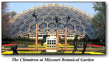
acknowledge that the principles set down by Buckminster Fuller for the construction
of geodesic domes were an inspiration for the development of their ideas.
We move now to 1962 when the still
often quoted paper of Caspar and Klug was published
in the Cold Spring Harbor Symposium volume of 1962. Some of the fundamental
ideas of the structure of icosahedral viruses were laid down at that time. They
discuss the concept of quasi-equivalence and
acknowledge that the principles set down by Buckminster Fuller for the construction
of geodesic domes were an inspiration for the development of their ideas.
The interest in and importance of this work may not have had many ramifications for virologists – diffraction patterns don't help explain how a virus can enter a cell or assemble into an infectious particle. Some of the questions being resolved by virologists at that same meeting included recombination in RNA viruses. The paper presented by George Hirst was "Genetic recombination with Newcastle Disease Virus, Polioviruses and Influenza". Recombination was not observed with Newcastle Disease virus. The structure of the RNA genome of influenza virus was not yet clear, but the high frequency of recombination led Hirst to suggest that "the RNA from this agent is often in fragments". The paper by Franklin and Baltimore ("Patterns of macromolecular synthesis in normal and virus-infected cells") discussed the possibility that a new RNA dependent RNA polymerase was induced in cells infected by Mengovirus and that the enzyme was coded by the viral genome. A significant number of the talks were concerned with transformation of cells by viruses and the characterization of those cells. In his concluding remarks Dulbecco stated a major problem of that period: "In the transformation caused by DNA-containing viruses the most outstanding event is the ultimate disappearance of the virus itself. It is most likely that the virus causing the transformation is never recoverable in infectious form from the cell it infected. This is an unfortunate event, because it renders extremely difficult if not impossible the isolation of viral mutants causing special types of transformation".
| Introduction | Some historical highlights: structural virology and virology |
| Solving the Structure of Icosahedral Plant Viruses | Picornavirus Structure | Poliovirus | Polio
The Influenza Virus Hemagglutinin | The Influenza Virus Neuraminidase |
Issues of Science and Society |contributors|
Home |