 |
|
|
|
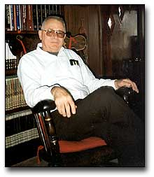 |
Roland Rueckert is featured here because of his seminal work on the structure of picornaviruses. As documented in Michael Rossmann's oral history, it was Roland that persuaded him to determine the rhinovirus structure. In addition to the work with Michael, Roland made a number of important contributions to our understanding of the replication and assembly of RNA viruses, particularly picornaviruses and nodaviruses. Here is a brief description, in Roland's own words, of his background:
"I was born on 24 November 1931 in Rhinelander Wisconsin, the first of four sons. My father, George, was unable to speak English when he immigrated from Germany in 1925. But he found a woman, Monica Seiberlich, "willing to take a chance on him" as he expressed it wistfully. He came to Rhinelander in 1930 to work in the grocery store of an uncle. We were poor, especially on things like clothing and shoes, but we never wanted for food and housing. Our family's big break came with World War II when, in 1942, my father hired out to work on the Alcan Highway needed for military transport to protect the Aleutian Islands under threat by the Japanese. One dollar per hour was a huge wage in those days; there was a bonus for those who stuck out the permafrost, bitter cold, biting insects and unrelenting work schedule - few did, but my father did! After a year of this my father returned with enough money to buy the store. That meant the entire family worked in the store, weekday evenings, all day Saturday and Sunday mornings. I began delivering groceries at age 13. It was illegal to drive at that age and I was very uncomfortable with it, but my father insisted and he was the boss. My first day out I almost hit a customer with the delivery truck! By God's good graces there was never another incident like that.
In sixth grade I won a Gilbert chemistry set, a reward for selling seeds for a commercial outfit, and realized almost immediately that I just loved chemistry. In seventh grade Civics class I wrote a vocational paper that some day I was going to be a chemist. I had the usual experiences with explosive mixtures and rockets. A concoction, of powdered magnesium, sulfur and potassium nitrate, once exploded in my face. I might well have been blinded had I not been wearing glasses; they were so peppered with embedded globules of molten magnesium I had to get new ones. On another occasion I showed a friend how to make nitrogen tri-iodide amine with ammonia and iodine. We wiped a spill off the floor with an old rag and hung it on a radiator to dry. That evening, when his mother picked up the rag it exploded in a puff of purple smoke covering her arm with brown iodine stains. She thought she was burned. Next morning I got a very angry lecture but she was unhurt thank heavens.
In 1949, I enrolled in the chemistry course, geared to professional chemists, at the University of Wisconsin. The campus was still overcrowded with students, veterans on GI Bill finishing their education and many of my classes were held in corrugated Quonset huts and so-called 'temporary buildings' a few of which survive to this day. I excelled in science and chemistry and every day was a joy because from there on I was free from the store.
By my junior year I had a job as a laboratory helper at McArdle Cancer Research Laboratory and after that was able to pay my own way through school. In 1953 with a BS in chemistry I was drafted into the Army as an enlisted man. After basic training at Fort Riley Kansas and Camp Pickett Virginia, they sent me to Walter Reed Army Medical School where I worked as an enlisted man on morphine and pentothal metabolism in a liver perfusion system. Our barracks, #73, included young scientists including Alan Campbell, the first to realize plasmids are circular, and Peter Guideschek so there was no shortage of stimulating companions.
I returned to the University of Wisconsin to earn a Ph.D. in oncology (1960) under the mentorship of Gerald Mueller. One of the papers from my thesis, on thymineless death in HeLa cells, became a citation classic (Cancer Research 1960) because it was a foundation for the technique used to select B-cells making monoclonal antibodies.
In 1960 NIH awarded me a three year postdoctoral fellowship to study virology in Germany. After a year with Wolfram Zillig at the Max Plank Institute in Munich, where I learned cell-free protein synthesis using bacteriophage phiXl74, I moved to Tubingen to learn animal virology with Werner Schaefer at the Max Planck Institute for Virus Research. He started me with influenza but I switched to mouse-Elberfeld virus as a better object for studying cell-free protein synthesis. I continued this work as a research virologist in Wendell Stanley's laboratory at the University of California at Berkeley (1962-1965) and later at the University of Wisconsin where I was hired as an assistant professor with a joint appointment in the Biophysics Laboratory and the Department of Biochemistry. The major problem during these five years was the inability to identify the protein subunit which we finally realized was being cleaved during assembly into four pieces (Rueckert, RR, Dunker, AK, and Stoltzfus, CM. Proc Natl Acad Sci 62: 912-919, 1969). It was not until several years later that we were able to unravel the complex process by which the protein subunit is synthesized and assembled."
Roland's early work established the underlying structural similarities among the different picornaviruses, which include poliovirus, rhinovirus - the common cold virus, foot and mouth disease virus, and others. As part of this work, he isolated the basic subassemblies of picornavirus capsids, a protomer containing four different proteins and a pentamer of five protomers. From these subassemblies and their interactions, he deduced the capsid assembly pathway, involving construction of progressively larger intermediates by sequential addition of distinct assembly domains, each made accessible in turn by prior assembly steps. He also helped to show that picornaviruses produce a giant polyprotein from which all viral proteins are cleaved by viral proteases, mapped many of the cleavages, and showed that major protein processing pathways were conserved among picornaviruses. His collaboration with Michael Rossmann and other colleagues, which resulted in defining the atomic structure of rhinovirus, was further extended to use the structure to determine important principles of virus inactivation by antibodies and drugs.
In an expansion of his studies on icosahedral RNA viruses, Roland initiated studies of Nodamura virus, a new virus able to infect vertebrates and insects. Subsequent work by his group defined Nodamura virus as the prototype of a new RNA virus family and established these viruses as valuable research models by finding conditions to grow them in cultured cells, showing their genome to be the smallest among animal RNA viruses, and elucidating their basic RNA replication, transcription and translation strategies. He defined the assembly-dependent proteolytic maturation pathway of their virion proteins and the position and structure of a nodavirus RNA encapsidation signal. In collaboration with Jack Johnson, he helped to determine the atomic structure of nodavirus virions, which has served as a foundation for numerous ongoing studies.
Roland ended his research in virology in 1996. Since that time he has become deeply involved in studies of forestry and ecology. He told us that his first timber stand improvement cutting, the result of two years planning, began in early 2000.
Roland submitted some reminiscences about his work with rhinoviruses and polioviruses to the Discussion section of our Web site. Those comments are reproduced here.
"Well, my first reaction to your history request was "That's a long time ago; I'm a different person now", meaning I'm not a virologist anymore. For three years I've been thinking and breathing forestry and ecology."
But he relented and continued:
"OK what's recorded below began as a short description, but as I typed it all came back pretty vividly.
One part of Michael's history is all wrong. We were not foggy about the protomer concept in 1985 - Michael was, but I wasn't. We published a paper in 1969 (The Structure of Mouse-Elberfeld Virus: A Model, R.R. Rueckert, A.K. Dunker and C.M. Stoltzfus, Proc. Natl. Acad. Sci. 62: 912-919, 1969) and a second paper in 1971 (Fragments Generated by pH Dissociation of ME-virus and their Relation to the Structure of the Virion, A.K. Dunker and R.R. Rueckert, J. Mol. Biol. 58: 217-235, 1971) proposing the protomer concept for picornaviruses. The only uncertainty was whether VP4 was still situated close by but, even there, the simplest possibility was that it was part of the protomer. The crystallographic structure settled it.
As for the relationship with Jim Hogle, you remember he was a student here with Sundaralingam. I had crystallized poliovirus by accident while post-docing with Wendall Stanley at Berkeley in 1964. Stanley made it clear that computing wasn't up to the task of determining a virus structure at that time. In 1978 when I learned Steve Harrison had solved the protein subunit structure of Tomato Bushy Stunt Virus I realized the time was ripe. I asked Sundaralingam, our only biological crystallographer at Wisconsin and himself crippled by polio. He was tempted but ultimately unwilling to make the necessary commitment. But Jim, then working on the structure of lysozyme, leapt at the opportunity. I encouraged him to do post-doctoral work in Steve's lab to learn virus crystallography; we'd send him poliovirus and if things looked promising maybe I could convince the Biochem Department to hire him with a joint appointment in our Institute (then called the Biophysics Laboratory). Because we planned to mail the virus, I suggested we'd be less liable to run into trouble with the post office if we used type 1 vaccine virus rather than Mahoney (Mahoney had been crystallized by Finch and Klug in 1960). Jim got crystals but they were very fragile in the X-ray beam. We tried lots of things to improve crystal stability, fruitlessly. I didn't want to risk sending mg quantities of polio through he mail. Finally, after considerable trepidation, I suggested to Jim he talk to David Baltimore about supplying him with Mahoney. That resulted in his collaboration with Marie Chow.
Within months of the time Jim got high quality crystals he received a very attractive offer from the Scripps Laboratory in La Jolla. I tried to convince the Biochem Department to hire Jim. Neither Biochemistry nor the Biophysics Laboratory at Wisconsin were able to marshal an adequate counteroffer and, to my keen disappointment, it became clear Hogle could hardly refuse the Scripps offer. I did arrange for Joseph Icenogle, a graduate student from my laboratory, to join Hogle at La Jolla and this enabled Jim to get off to a fast start at Scripps. (Icenogle later joined the virology section at the Centers for Disease Control in Atlanta.)
Meanwhile at the Strasbourg Virology Congress in 1981 Michael Rossmann, having learned of Hogle's success, told me that he planned to proceed vigorously with poliovirus crystallography. In Michael's hotel room that evening I argued against this plan on the grounds that it might jeopardize Hogle's research career and that it made better sense to study a picornavirus belonging to a different family and thereby benefit from the comparisons that would emerge as the structures were complete. I suggested human rhinovirus 14. I knew it could be produced and purified in sufficient bulk, was stable enough for crystallization and posed no health hazard to lab workers. The latter point ultimately proved crucial in a race between Hogle and Rossmann to solve the first crystallographic structure of an animal virus. I agreed to provide virus for preliminary studies and totrain key personnel in Rossmann's laboratory with skills necessary for growing and purifying virus. To promote communication and maintain momentum we initiated regular annual meetings at Wisconsin.
I was interested in correlating picornavirus structure with its functions. How did they attach to cells and what kind of structural rearrangements are involved in the infection step, i.e. release of its RNA genome from the shell into the cytoplasm of the host cell? I hoped that neutralizing monoclonal antibodies might provide a key tool. That antibodies might be useful for this purpose was first suggested by Dulbecco and coworkers in their 1956 paper on neutralization of animal viruses. They interpreted kinetic evidence, much like that used in radiation target theory, to mean that a single antibody molecule could neutralize a poliovirus particle. Moreover they argued this was not at an attachment step. But because of the complexity of antibodies in antiserum, purification of the antibodies in question was technically infeasible. The arrival of monoclonal antibody technology changed all that. Inspired by Phil Minor's preliminary work on neutralization resistant mutants of poliovirus, we launched a program aimed at defining neutralization sites on HRV14.
Ann Mosser began with poliovirus and then taught Barbara Sherry how to make neutralizing monoclonal antibodies against HRV14. Barbara eventually isolated 35 B-cell clones producing neutralizing antibodies. These she used to isolate neutralization-resistant ("escape") mutants. These mutants were then used to classify the panel of antibodies into four groups using a microtiter neutralization assay. Sequencing the RNA showed that most of the escape mutants differed by substitution of one amino acid for a patch at the surface of the virus. Moreover each patch was correlated with one of the four antigenic groups previously defined in a novel way by screening for cross-resistance.
Within a month of the time Barbara located the four escape mutation patterns, Rossmann and his coworkers had solved the rhinovirus structure to a resolution good enough to see that at least two of the mutable residues resided on the surface of the protein shell. He invited us to bring our collection of mutations to Purdue and help them trace their location in the electron density map. The map consisted of over a hundred planes stacked in a pile some 15 inches high. We color coded each alpha carbon in the chain, blue for VP1, green for VP2 and VP4, and red for VP3. It was a tedious process - and a tense one! One by one, over the course of a long day, we identified one after another of Barbara's mutants. At the end our hearts jumped for joy. Every single one of the escape mutations was located at the surface of the shell. Moreover the mutations were organized in four patches with each mutable side chain projecting into the patch to which it had been assigned by the cross-resistance mapping. No exceptions. It was an incredible high - total intoxicating elation. It gave us an unanticipated conviction of certainty in our results that one almost never experiences in experimental science.
I realized at this moment the privilege of working with Michael Rossmann - and the luck of it. Michael accepted the disadvantages of working on the rhinovirus project, thus protecting Hogle's career. Poliovirus was known to form crystals of high quality, its sequence was known and antibody resistant mutants were already accessible. None of these advantages were available for HRV14. Yet Michael conquered these disadvantages, including a late start by his daring, determination and skill. Oh, and the element of luck? Rossmann was able to use the synchrotron to collect diffraction data while Hogle was not. That is because HRV14, unlike the Mahoney strain of poliovirus, was not considered a safety hazard. That, in the end, may have been the clinching factor in that exciting race for the first animal virus structure.
| Introduction
| Some historical highlights: structural virology
and virology |
| Solving the Structure of Icosahedral Plant Viruses
| Picornavirus Structure | Poliovirus
| Polio
The Influenza Virus Hemagglutinin | The
Influenza Virus Neuraminidase
| Issues of Science and Society |
contributors| Home |