 |
|
|
|
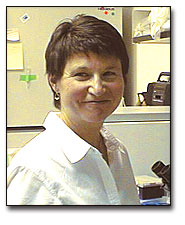 |
Judy White is currently a Professor of Cell Biology at the University of Virginia. We asked her to tell us about some of her remembrances of the time that the structure of hemagglutinin was solved and how that influenced her own research on deciphering the mechanism of fusion. Her narrative follows:
The beautiful HA structure, so expertly solved by Don Wiley, Ian Wilson, and John Skehel, has been a part of my professional life right from its inception. Even the anticipation of the structure played a major part in guiding my research interests.
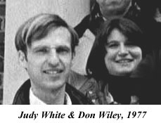
I have the honor of having been Don Wiley's first graduate student. Don and I had dreams of solving the structure of the Sendai virus fusion protein and, more importantly (from my point of view), of solving the "fusion problem" - just how do you get two membranes to fuse so that, for example, a virus can inject its nucleic acid into a cell to start the replication cycle.
|
My recollection is that it was a lecture by Steve Harrison, in a structural molecular biology course at Harvard co-taught by Don, that sparked my interest in the basic problem. The lecture was about the structure of bacteriophages. As part of the lecture, Steve discussed how the phage injects its DNA into a bacterial cell. I thought this is really cool; it's a little spacecraft machine that sits down on a cell, docks closely to the bacterial membrane, and then inserts its infectious material. |
But how does an enveloped animal virus do this? The observations that Sendai virus caused red blood cells to fuse and could induce giant syncytia in tissue cultures suggested the simplest mechanism in which the enveloped virus would fuse its membrane with that of the host cell and in so doing (without lysing the cell) would provide a tidy way to get its nucleic acid into the host cell.
The general topics of virus mediated cell-cell fusion and virus-cell fusion combined my growing interests in virology and membrane biology. I had a seed of an interest in virology because a cousin was stricken with polio. I was interested in membranes from my undergraduate days, from coursework and several research experiences. I actually started a Ph.D. thesis at Harvard with Guido Guidotti working on a protein that underlies the red blood cell membrane. The project was going well and Guido was a fantastic advisor but I had been bit by the virus bug and I really thought it would be a wonderful experience to work with Don (I had done a rotation in his lab). Don was a new professor full of energy and ideas and enthusiasm. So, I switched labs. (Dr. Guidotti was very supportive.)
I was working on Sendai virus, studying the biology of the fusion reaction, purifying, characterizing, and reconstituting the Sendai fusion (F) protein, and trying every protease in the book to try to isolate the ectodomain of the protein for crystallization (by analogy to the bromelain released fragment of HA, BHA). We never got an intact oligomeric ectodomain of the Sendai F protein. Nobody has, to this day. So, I did a lot of basic protein chemical characterization of the Sendai F protein and reconstituted some of its biological activities (e.g., hemolytic activity). A significant part of my thesis was working with Mary-Jane Gething [at the Imperial Cancer Research Fund (ICRF) in London, England] on a large-scale purification of the Sendai F protein and obtaining its N-terminal sequence. And, as described by Don and John, that N-terminal sequence was really impressive! It was so much like the sequence at the N-terminus of HA2 that it absolutely convinced me (I believe all of us: Mary Jane, Don, John, Mike Waterfield) that if Sendai could cause fusion, then so could flu. We just didn't know the missing parameter yet (low pH); more on that later. In retrospect, another important part of my thesis is a chapter full of what are largely negative results. The effort was an attempt to get BHA to bind to detergent micelles or phospholipids. Given their hydrophobic character we reasoned that these N-terminal "fusion peptides" should love lipid and detergent. I assayed for binding of TX-100 (tritium labeled) to BHA; no binding. I treated BHA with up to 2% SDS at room temperature and 0.5% SDS at 37 degrees and saw no binding. I tried binding of a monomeric lecithin to BHA, no binding. (All of these experiments were done at neutral pH). In an equilibrium gel filtration experiment, however, I did see a small peak of micellar lipid binding to BHA. We never made much of that, never published it. It may have represented binding to a small fraction of BHA that had changed conformation at the micellar lipid interface.
You asked me what the lab was like. It was at the same time both quiet (for me as a biochemist) and exciting. I would come in pretty early in the morning and usually have the place to myself. The only other person working in the wet biochemistry lab at the time was Micheline McCarthy who was a graduate student with Steve Harrison.
|
The crystallographers usually came in later and worked across the hall. Don spent most of his time, of course, in the crystallography side of the lab. But every once in a while he'd pop back into the biochemistry lab. My sense was that solving the HA structure was simultaneously stressful and exciting. They had crystals for quite some time and were methodically and (to me) painstakingly taking lots and lots and lots of pictures and analyzing lots and lots and lots of data. From my perspective Don and Ian (Wilson) had tremendous perseverance. One knew the structure was important and that it was bound to be interesting, but their drive saw it through. |
 |
By the way, I was thrilled to see the picture of the BHA crystal decorated with red blood cells on the website. I remember looking down the microscope in the tissue culture lab (in the basement) at those crystals. It was so exciting! It was also a fun time in the Gibbs lab. During my time in the lab some other notables (notable scientists and notable characters) worked with Don and Steve: Ian Wilson, Clarence Schutt, Art Olson, Jim Hogle, and Pamela Bjorkman (my last year). (We had some great bike rides and some great meals!)
My recollection is that the decision for me to go to London just evolved. We had heard that Mary-Jane Gething was also working on purifying Sendai F and somehow an arrangement was made for me to go to London to work with her. And so began a wonderful collaboration and friendship. London was wonderful! The lab (Mike Waterfield's) was wonderful! And the whole experience of being abroad and working in a "foreign" lab was wonderful! Mike's lab at the ICRF was a well-equipped protein chemistry lab with loads of columns and fraction collectors and protein sequencers. (This does mean protein sequencers, sequencing of nucleic acids to determine the amino acid sequence of a protein had not yet taken over.) It was bustling - in contrast to my quiet biochemistry lab in Gibbs. We also had loads of starting virus for purification. Virus preps (in eggs) were done on a much larger scale than we did in Don's lab. That was eye opening. Periodically I'd go to Mill Hill to see Don and John. It was usually timed for an egg prep day. There were thousands of eggs, trays stacked high. Whoever came into the lab got an egg punch or helped out in some way. It was quite a production.
The most important question you asked was whether the structure influenced my thinking and the course of my work. The structure of BHA (and the subsequent structure of TBHA2) MOST CERTAINLY DID!! I was fusion-focused in viewing the structure. We knew that the HA2 NH2-terminal fusion peptide was going to be important (confirmed by later mutational analysis). Although I didn't expect the fusion peptide to be on the surface of HA, I, at least, expected it to be near the top (nearer the target than the viral membrane) of HA. Yet, it is closer to the viral membrane end of the protein (tucked in). This suggested that the protein would have to undergo a large conformational change, to expose the fusion peptide and to get it to the target membrane. At that point there were two big questions (in my opinion): the nature of the conformational change(s) and the trigger - what induced the conformational changes.
Before the structure was published (1981) I had moved to the European Molecular Biology Laboratory in Heidelberg to work with Ari Helenius on virus fusion and the cell biology of virus entry. Ari was working with Semliki Forest virus (SFV). During my first months in Heidelberg, Ari and Kai Simons (and co-workers) were finishing up what has become a classic paper. It was published in early 1980 (Helenius, A., J. Kartenbeck, K. Simons, and E. Fries. On the entry of Semliki Forest virus into BHK-21 cells. J. Cell Biol. 84: 404-420). This paper showed, for the first time, that low pH could induce virus fusion, in this case SFV to liposomes. Most importantly and uniquely, the Helenius paper showed that low pH is a physiologically relevant fusion-inducing trigger: the virus enters cells through endosomes which maintain a mildly acidic pH. In my first year in Ari's lab, I basically refined and characterized in detail the SFV-liposome fusion reaction. Doing experiments with your own hands makes you a believer and I quickly became a believer in low pH as a fusion-inducer. The pH profiles were sharp and highly reproducible. Unlike Sendai, SFV had never been shown to induce syncytia. We (I truly don't remember whose idea it was) then put two and two together and asked if SFV would induce syncytia in tissue culture cells if we acidified the culture medium - a very simple experiment. And, voila, giant syncytia! Quickly on the heels of that experiment I did the same thing with influenza virus as well as with vesicular stomatitis virus (VSV). And, sure enough both viruses induced massive syncytia, but only at low pH (White, J., K. Matlin, and A. Helenius. J. Cell Bio. 89: 674-679, 1981). Around the same time, Maeda and Ohnishi (FEBS Lett.122: 283-287,1980) showed that influenza could induce hemolysis and fusion of red blood cells (RBCs) and John Lenard (Lenard, J. and D. Miller, Virology, 110:479-482, 1981) showed that SFV and VSV could induce RBC hemolysis at low pH.
Now fully believing that low pH induces influenza virus fusion, I reasoned that I finally knew how to get BHA to bind to liposomes. So, soon after I saw flu-induced syncytia (at low pH) in the microscope, I asked John Skehel to send me some BHA (to Heidelberg), which he did. (I truly didn't know what experiments John was doing or was planning to do.) When I got the BHA, I mixed it with liposomes, treated one sample at low pH and then ran flotation gradients (the ones referred to by John in his oral history). I analyzed the gradients for protein and lipid. In, and only in, the gradient of the low pH treated sample, BHA was in the same fractions as the liposomes. That simple quick experiment provided one of the highest highs of my scientific career. (I vividly remember calling John to tell him the results from our lab in the EMBL in Heidelberg.) The flotation gradient experiment is in the 1982 PNAS paper (Skehel, J.J., P.M. Bayley, E.B. Brown, S.R. Martin, M.D. Waterfield, J.M. White, I.A. Wilson and D.C. Wiley. Proc. Natl. Acad. Scie 79: 968-972, 1982) and I think it was pretty important; it proved that the HA ectodomain could convert from a hydrophilic to a hydrophobic entity that could stick to a target membrane simply by a brief exposure to low pH.
For me the next years were full of further biochemical and immunological analyses of the conformational changes in HA and making and characterizing HA mutants for fusion phenotypes. These activities were directly motivated by the BHA crystal structure. Two other activities (on HA projects) included demonstrating that HA (expressed in tissue culture cells) was necessary and sufficient for low pH induced fusion and analyzing the membrane dynamics during fusion: breaking down the process into target membrane binding, hemifusion, and fusion pore stages.
Then came the next big jolt - the structure of TBHA2 (the core of low pH treated BHA)! By that time we knew about many conformational changes in HA, for example separation of the globular head domains and exposure of the fusion peptides. There were also hints in the literature (from Ward and Dopheide and from Peter Kim) that the so-called B-loop wanted to be a helix. (I too was told that the BHA structure was "likely wrong" in the "B" region, I believe by Jeff Stock) But I certainly didn't anticipate the two other big changes -the helix to loop transition that jackknifes the HA2 D helix antiparallel to the C helix, and the extension at the C-terminal end of HA2, the "leash" between the core domain and the transmembrane domain. Based on these changes we proposed a model in 1995, which we continue to test, in which (as yet hypothetical) intermediates between the BHA and TBHA2 states drive distinct stages of the fusion process: target membrane binding, hemifusion, fusion pore opening. The latest version of this model can be accessed via my lab homepage.
So, it's almost 20 years post the BHA structure and we are still doing experiments on HA! From my vantage point the crystal structures are just the beginning! I, and I am sure, numerous others are therefore deeply indebted to Don Wiley and John Skehel (and their collaborators) for illuminating the structures and mechanism of a fascinating protein that continues to entertain us.
| Introduction
| Some historical highlights: structural virology
and virology |
| Solving the Structure of Icosahedral Plant Viruses
| Picornavirus Structure | Poliovirus
| Polio
The Influenza Virus Hemagglutinin | The
Influenza Virus Neuraminidase
| Issues of Science and Society |
contributors| Home |