 |
|
|
February 26, 27, 1999 Recorded and edited by |
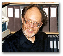 |
Michael Rossmann is the Hanley Distinguished Professor of Biological Sciences at Purdue University. He was born in Frankfurt/Main, Germany in 1930 but he and his Mother left Germany for England in 1939. His early scientific education was at the University of London where he studied physics and mathematics and received a Master's degree. He moved to Glasgow in 1953 where he taught physics in the technical college and worked on his Ph.D. He attributes his initial interest in crystallography to Kathleen Lonsdale and to a book on the subject by W C.Bunn. In her biographical memoir of Kathleen Lonsdale, Dorothy Hodgkin wrote "She helped to start the Young Scientists' section of the British Association ('Never refuse an opportunity to speak at schools' was one of the memorandum notes she left for herself.) Michael Rossmann was one of the schoolboys who heard her and became a crystallographer as a consequence" (Biographical Memoirs of Fellows of the Royal Society 21: 447-484, 1976).
Michael told me that the quote was not exactly correct. Kathleen Lonsdale was on the board of governors of the high school he attended (a Quaker school and she was a prominent Quaker). She gave the school a few tickets for the lucky ones to attend the annual Christmas lectures at the Royal Institution for high school students. That year they were given by the astronomer Royal. But it was in this way that he got to know Kathleen who took the students around the basement of the Royal Institution where Michael Faraday used to work. Later Michael turned to Katleen Lonsdale for help to find a research lab and that is when he started reading the book by Bunn.
Michael began his career as a crystallographer when he became a student of J. Monteath Robertson at the University of Glasgow and received a Ph.D. from that University in 1956 in Chemical Crystallography. The title of his thesis was "A Study of Some Organic Crystal Structures".
In 1956 he and his family (which at that time included his wife and two children) moved to the University of Minnesota where he worked for two years with Professor William Lipscomb publishing several papers on the structure of terpenoids and writing computer programs for analyzing structures. He then returned to England and to Cambridge where he worked with Max Perutz on the structure of hemoglobin.
The first part of the interview covered his early work on x-ray crystallography and
hemoglobin and then I asked:
SS How did you get interested in viruses?
MR Actually in 1960 the Meeting of the International Union of Crystallography, a meeting that happens every 3 years, was held in Cambridge and I went to that meeting. The ideas that are now known as molecular replacement came to me during that meeting. I realized that we should be able to recognize the relationship between 2 similar chains in hemoglobin without any heavy atoms. This calculation is known as a rotation function today. There were actually 3 stages to the idea. At that time I realized one should be able to work out a complete structure (without heavy atoms) based on the symmetry between the alpha and beta chains (of hemoglobin) which I called non-crystallographic symmetry. And I thought I could work all this out by Christmas. It was August or sometime in the summer when I attended that meeting. It took another 25 years! I did get done by Christmas the rotation function calculation. I programmed it and worked out the many consequences of it and showed one could do this.
In today's crystallography there are only a few well known methods for calculating structures. There's isomorphous replacement which is Max's idea, molecular replacement, and multiple-wavelength anomalous dispersion which is now called MAD by W. Hendrickson and Janet Smith. What else? That's it! These are the methods in modern protein crystallography and molecular replacement is particular useful if you already have a similar structure. It's also useful if you have many identical units so David Blow and I published the first paper on molecular replacement in 1962 but the ideas started in 1960. (David Blow had been a graduate student of Max's and had worked on hemoglobin for his Ph.D. He spent 2 years with Alex Rich, first at NIH and then he moved with Alex to MIT for his 2nd year. When he came back to Cambridge he had a desk almost next door to Michael's and they published several papers together.)
I immediately recognized the value of the concept of molecular replacement to viruses.
In a 1962 paper on the subject (M.G. Rossmann and D. M. Blow Acta Cryst. 15:24,1962) the authors wrote: " Evidence is accumulating that many of the larger protein molecules are made up of identical or closely similar subunits. The reasons for expecting this for the protein part of virus structures was set out by Crick and Watson (1956) and the prediction has been amply confirmed in crystallographic studies of spherical viruses."
MR So this was the first paper. This is an application to hemoglobin and this is hemoglobin which shows a peak which is the right place for showing the alpha chain and the beta chain are the same. And I realized that this was going to be important for viruses.
SS So you had seen the relationship between molecular replacement and viruses but when did you begin to think about working on viruses, yourself?
MR Right there and then. I am doing a review of a National Science Foundation Grant proposal. I wrote my first grant proposal about a year before I came to Purdue so I would have some money when I arrived and I want you to see the name of it. It's "Proteins and Viruses". I came to Purdue in '64 and must have written this first grant proposal in 1963. Roger Burnett was my first graduate student here and the first project he worked on was to crystallize a plant virus, probably CCMV (cowpea chlorotic mottle virus). One of the people in the neighboring department was John Bancroft, a plant virologist. I had Roger working with John trying to crystallize CCMV, but he didn't succeed.
In Cambridge I was having a wonderful time - doing all the things I liked to do and not getting any real results. I was developing techniques. I realized that when I was coming to Purdue if I wanted to be successful I had to get some real results. The big project I worked on - do you know anything about the work I did on dehydrogenases?
SS Yes
MR You know everything.
SS Well, we've both been around for a long time
MR But you're a virologist.
SS No, I was trained as a biochemist
MR Oh I see OK. So for the last half year or so in Cambridge I was learning how to extract lactic dehydrogenase from pig muscles. I was going to a slaughter house collecting material.
SS You were becoming a biochemist.
MR Yes I had to do this and I continued that here because we worked on LDH. We actually did dogfish LDH. Eventually we had to purify it.
SS Do you remember why you picked the dehydrogenases?
MR Oh yes very much so. I was looking for something which would have like hemoglobin - relationships - and at that time (Clem) Markert had just showed that LDH had isozymes. And so it occurred to me that the different isozymes might have similar structures - the heart and the muscle form. They might be basically like the alpha and beta chains of hemoglobin. Also I was looking for something for which there was evidence that it could be crystallized and LDH had been sort of crystallized and I was looking for something which would have sulfhydryl groups for heavy atoms. LDH had a lot of work done by Professor Pfleiderer in Germany on LDH investigating sulfhydryl groups and I was looking for something which would have symmetry so I could use the ideas of molecular replacement.
SS Was that considered at that time to be a big jump? Was it considered a difficult project?
MR Oh yes, when we actually solved it, it was by far the biggest structure that had been solved to date.
SS What had been done? hemoglobin, myoglobin and lysozyme.
MR By the time we actually solved LDH, more than that. By that time myoglobin, hemoglobin, lysozyme, chymotrypsin. I was working on chymotrypsin with David Blow.
SS Was LDH the largest enzyme?
MR Yes, by far, a factor of 4 maybe and it was also a subunit enzyme. Hemoglobin was the only other structure that had that property. So I very carefully selected something which I thought would be interesting for all these accounts. In one of the early papers on LDH, I go through the ideas of why I chose LDH. So, LDH was successful. I then decided I must pursue viruses and so I took a sabbatical. I had been here (Purdue) for 6 years.
Even before I went on my sabbatical I worked with Peter Gilham here. I started to isolate Qb phage) but it was very impure and I didn't make any progress. Then I went on sabbatical to work with Bror Strandberg who I had gotten to know in Cambridge when he was working with John Kendrew.
SS What year was this?
MR 1971
SS So you went to Sweden to try to work on a virus?
MR Yes and I did. I worked on satellite tobacco necrosis virus. One of the things I did there was to run a rotation function which should and did show the orientation of the virus in the crystal itself.
SS So the virus had already been crystallized?
MR Oh yes. It has been crystallized in the late 30's and Dorothy (Hodgkin) and Bernal had taken some X-ray photographs but they had not continued. Bror was busy propagating the virus on plants and isolating virus and he had a nice setup for doing all that.
In 1944 Dorothy Crowfoot (Hodgkin) and G.M.J. Schmidt obtained some crystals of this virus from N. W. Pirie. They took some X-ray measurements before the crystals deteriorated and thought that their pictures were so "remarkable" that they published their findings and preliminary calculations (See the History Time Line, 1945).
SS I'm trying to imagine when people first starting doing this, whether they really thought it was going to be possible.
MR No, No. I remember about this time, no it was later in 1976 - at a meeting in Italy, in Sicily at a place called Erice. I was the first director of the crystallographic school there. They have one every six years now and I was at the first one. I remember sitting on an old Roman temple stone talking with David Phillips and David was saying "you really shouldn't be growing viruses. You should be working on enzymes and enzymes do things fast, viruses are quite dead. Why do you do this?" So we had a discussion about this and that was the general atmosphere.
SS Let me go back a minute to ask: Were you influenced at all by the paper of Caspar and Klug in 1962?
MR Yes-I mean it was very important. At that time people were saying it was not worthwhile looking at a T=1 virus because everything is obvious, like satellite tobacco necrosis virus where T=1. Yes, one of the first things I did when I went on sabbatical was to get the Cold Spring Harbor paper of Caspar and Klug and I read it very carefully.
SS It is a classic paper.
MR Yes. It is. I quite agree. Very classic, very interesting. Just yesterday one of Tim Baker's students had a Ph.D. committee meeting and I was on it and I pointed out that what he had was not at all the classical Caspar and Klug lattice and I don't believe anybody even realized that.
SS What virus was he looking at?
MR This was an iridovirus, (a double-stranded DNA virus) a very, very big virus, about 2000 angstroms (in diameter).
SS So now we're back to the 70's and you are in Sweden..
MR I ran the rotation function and got an orientation, but the trouble was this rotation function looked as though the virus was octahedral. If it were octahedral, there were only 24 parts rather than 60 parts. So Bror "stood on his head" and got people to do sulfhydryl determinations and molecular weight determinations and everything turned out to agree that it looked like it was octahedral. Then there was a Cold Spring Harbor meeting in the summer of 1971. It was a very important meeting for other reasons, the PDB, the protein data bank, started there. Bror gave a talk, and he had hardly opened his mouth when Aaron Klug who was on the front bench started heckling him (saying) "It was impossible, viruses are icosahedral. It was obvious, you must be wrong". Then there was a general backlash against the rotation function,
In particular Don Caspar said that it was very important to look at viruses. What Don had done with tomato bushy stunt virus - he had done this in Cambridge - was to look at what he called spikes - spikes of intensities in that very famous paper of his in Nature - and he said "that's the way to look at the symmetry. It's only a primitive way, rotation function. And so Aaron was quite emphatic that this was wrong, and of course it was wrong. But it was also actually right. It turned out that satellite tobacco necrosis virus had crystallized in a very special way where one of its icosahedral 2-fold axis was at 45 degrees with respect to the crystallographic 2-fold screw axis. So these two-45 degrees were at 90 degrees apart and that gave rise to octahedral symmetry. So the octahedral symmetry peaks were actually twice as high as the icosahedral peaks which were in the background so they were swamped by the bigger, larger octahedral peaks.
SS Who figured that out?
MR Well, in a sense, Aaron figured it out. After the meeting he wanted all the data in Cambridge and he and John Finch looked at it. But I also figured it out soon after I got back to Sweden. I was on sabbatical there and realized the mistake but wouldn't have realized it without Aaron's pushing. I realized that there were really two of these viruses and they must have separate peaks and not the same peaks and it must be right what Aaron said. The rotation function was right. It gave basically the right answer. What was wrong was our interpretation. And the unfortunate thing was all the biochemists were pushing their results to agree with what we had said. Everything seemed to be agreeing but really the biochemistry was wrong.
SS What kind of biochemistry data was it?
MR The number of sulfhydryls in the virus - the molecular weight of the subunits - compared with the molecular weight of the whole virus and I think there were some other data.
SS So the biochemical data were not good enough to really help you.
MR It was deceptive. To me of course it was very very depressing. The fight with Aaron Klug gave me a very nasty taste at the time. [Michael goes to get some papers] "Structure of viruses" oh yes- here we look at this. This was our paper here which we submitted. Wonderful pictures! They are all precession photographs, we wouldn't do that today. Here are all these rotation functions. Here is what we submitted, which was the original manuscript. Then after the meeting (a Post-Symposium Discussion) "Interpretation of the rotational function map of satellite tobacco necrosis virus: octahedral packing of icosahedral particles". That's what Aaron Klug said. Then also additional notes by Akervall et al. There we give the real interpretation of the rotation function.
This is an interesting paper (He is talking about the next paper in the volume) - the first image reconstruction of a spherical virus- this is all negative staining.
SS One of the things I was trying to think through was when we talked earlier about seeing the structure of hemoglobin, but I was thinking of the viruses was: What was it like to first see the structure after you had the maps? Did you know what you were going to see based on the image reconstructions?
MR Absolutely not
SS Didn't the electron microscope give you an idea?
MR No, no, the electron microscope didn't give us any idea. Well, for tomato bushy stunt virus Steve (Harrison) probably had a poor reconstruction and it might have helped him get the phases. I can't remember. But this was all crystallographic stuff. The electron microscopy didn't really get into swing until cryo-electron microscopy came along. Now Aaron Klug got the Nobel award for his ideas on image reconstruction from EM and that must have been in the early 80's when Aaron got the Nobel (1982). Yes, because I remember it being announced when I was in Hamburg collecting data on rhinovirus. Then the cryo-EM didn't get started until maybe '85 or '86.
SS When you came back (from sabbatical) was that when you began on viruses?
MR Well, I had started on Qb but I realized that was probably not the right thing and started working on plant viruses and another plant virologist was in this department, Richard Lister and through him I got the idea of working on southern bean mosaic virus.
About that time, my first post doc who was willing to work on viruses - I came back from Sabbatical in the summer or Fall of 71 and then Jack Johnson - who was a graduate student with Bob Jacobson with whom I worked with in Bill Lipscomb's lab - Jack came to the lab about 2 months later by which time I had started with southern bean mosaic virus and Jack then took over the preparation of SBMV and the crystallization of it. This was the first post doc I had working on viruses and he did very well
SS So I'm trying to get you to think about that time and whether you were worried whether it would work.
MR I never thought it would not work. With Bror, you know, I had learned that it was possible to collect data. Now one thing I changed from Bror's lab, he was doing all this with what we called precession photographs and I realized that's no good for a larger virus and there's another technique which was just starting to be developed by Alan Wonacott, one of my former post docs. It was called oscillation technique, which is the best way of just collecting data from day to day and in 1973, maybe, there was a conference in Groningen, organized by the company that was manufacturing the first of these oscillation cameras,to which I actually asked Jack Johnson to go. Jack went to that meeting and I had a post doc Geoffrey Ford an Englishman who I had asked to write the necessary software to analyze the oscillation photographs, but that didn't work out. Geoffrey eventually left and went back to England. I realized I had to do it myself.
SS I was going to ask if it was difficult to find post docs who were interested in this project? Obviously, Jack thought this was worth the time and effort.
MR Yes, In retrospect, I think he was quite foolish.
SS In retrospect he wasn't.
[Both laugh]
MR It was very risky shall we say.
SS It was probably a different time that people felt that they could afford to be more risky.
MR I wonder, I don't know. He was the only postdoc I had for a long time on that.
SS So all the other people came after Jack?
MR Yes, Ivan Rayment came, Andrew Leslie, and Tsukihara came from Japan.
SS Let's go back to the questions I was asking about, there are two questions. What was it like to see the structure? And then I read not only your Nature paper but Steve (Harrison) had a "News and Views" afterwards titled "virus crystallography comes of age" and he makes the point that it was a surprise that they (tomato bushy stunt and southern bean mosaic virus) were so similar and I wanted to discuss that.
MR Andrew Leslie was really the critical person on that. I guess it was in the summer of 79 and he had arranged to go back to England, to Imperial College, actually to be a post doc in David Blow's lab. So big efforts were made to solve the structure for southern bean mosaic virus. We added more and more heavy atoms to get derivatives. The map just didn't look right. So I'll tell you the answer right away. It turns out the program for averaging had been written by Jack, and the input of this requires accurate cell dimensions and if your cell dimensions are not accurate the averaging doesn't work because say you shrink one axis and you rotate this point to that point, you are going to compare it in the wrong place. We kept on using the same input file and the input file had some old inaccurate cell dimensions and so it turned out that we had used too inaccurate cell dimensions and we had much better cell dimensions than we actually used in the first place. So, later after Andrew left and after we had actually interpreted the map we recognized this and Tsukihara recognized this and then we were able to get a much better map. Abad-Zapatero did that, a really beautiful map. We probably could have solved southern bean mosaic virus 2 years earlier had it not been for this mistake. We kept on adding more heavy atom derivatives. Now, I said Andrew left in the summer of '79. He was determined to solve the structure. Was it Andrew or was it me? I think it was maybe the combination of the two of us, I can't remember. By this time, Steve had the tomato bushy stunt virus maybe a year earlier and I think we had the coordinates from Steve. This is a beta barrel and you have these beta strands and one of the deficiencies in our maps - we couldn't resolve very readily one beta strand from the next - although the ends were very obvious and I think, maybe Andrew recognized that he really had a beta barrel there and now the question was – did we have the same topology? After awhile we recognized we had the same topology, but it was a very poor map. We couldn't have recognized the structure without Steve having solved the structure of tomato bushy stunt virus but - here we are - we have the same structure. It's clear we have the same structure but we wouldn't have known that, we wouldn't have been able to determine that without the knowledge from Steve. As I said a half-year later we had a much, much better map because we recognized the reason why we had had a poor map. So it was a big surprise. We didn't expect this, but it was wonderful because it gave a lot of meaning to solving these virus structures.
SS Once you solved the structure what did it make you think about? You had the structure, what does this tell you about function?
MR Well, it showed us something about evolution. When we actually did have a good map we didn't have a sequence of southern bean mosaic virus. I persuaded Mark Hermodson who was Head of Biochemistry to determine peptide sequences and we fitted the peptides into the map but this was after we had the much better map. By this time, it was 1980, and I took my next sabbatical. I took it in Cambridge with Richard Henderson to learn some electron microscopy and this was just at the time when computer graphics was coming in. I had invited Alwyn Jones, he is the king of computer graphics, crystallographic computer graphics, who had done his post doc work in Germany with Robert Huber, later a Nobel laureate. By that time he had gone to Sweden to Upsula where I had been on sabbatical - he still is there now - and I invited Alwyn to come to Purdue. I had bought from Washington University a homemade computer called MNX. It was a very nice computer and Alwyn installed his early Frodo program. This was the first useful graphics program. There were lots of people programming computer graphics including us.
This is in the late 70's. And then around Christmas 1980, I came back from Cambridge from my sabbatical and Alwyn arrived here and stayed with us a few weeks. He didn't finish this installation. I finished the installation of the computer graphics and with that computer graphics, we were then able to look at southern bean mosaic virus which I did while Mark Hermodson was doing the sequencing of peptides and we were able to fit the peptides in while Cele Abad-Zapatero helped with the peptides. He was building the model, I was doing the graphics.
SS. Let's spend a little more time on southern bean mosaic virus, because that was really the first time you really had a structure to look at.
MR It was the second virus structure.
SS What you said was that it told you more about evolution than about function.
MR It told us about evolution but then when I started to learn the graphics - I spent a half a year studying the graphics - I worked out all the structural relationships and I started to understand how - what Steve Harrison called - the divided contacts, work; how the "AB" contacts and the "CC" contacts work, how the C subunit is the more ordered structure. The polypeptide is there between the two C subunits and divides them and makes the icosahedral 2-fold axis. The AB subunit contacts are between the quasi-two fold axis, there is a tilt there. And I worked all this out and of course this was published in a paper with Mark Hermodson and others. There I go into great detail about the way the T=3 symmetry works. And the T=3 symmetry doesn't actually work quite like Caspar and Klug predicted, but it roughly works. There are deviations and what is accurate and what is inaccurate, what is quasi and what is not quasi. I worked all this out for southern bean mosaic virus. I think it told us a lot about quasi-symmetry and T numbers and so on, that was very interesting. We also did some things on calcium binding and that was interesting.
About that time, I had to renew my NIH grant. NIH was very clear about directing me toward animal viruses and in 1980 I went to the Strasbourg International Conference of Virology. I had the opportunity to talk with Roland Rueckert . We were not that far away each other in America but we had to go all the way to France to talk and we decided to combine our projects on rhinovirus. Actually, even before, Aaron Klug had shown that poliovirus crystallizes and so I had Sherin Abdel-Meguid, a post doc with me go to Ellie Ehrenfeld's lab in Salt Lake City to start working on poliovirus. Of course Jim Hogle was working on poliovirus too and so Roland said you really shouldn't compete like that. Roland was quite right because we had no idea or very little idea that rhino and polio viruses would be so similar.
SS Maybe this is really hindsight but since you had just found that southern bean mosaic virus and tomato bushy stunt virus were similar, it couldn't have been quite as surprising to expect polio and rhino to be similar.
MR No, No, in fact, when we did solve rhino virus - it is very, very similar to southern bean mosaic virus - the only difference is that southern bean and tomato bushy stunt have 3 identical subunits A, B and C. In the picornaviruses A is VP1, B is VP3 and C is VP2.
SS I would have thought you would have expected rhino and polio to be similar.
MR No. we really didn't. But something did happen. Our first crystals, not very good, of rhino virus were actually pseudo-isomorphous to Aaron Klug's crystals of polio. Then we realized, although we could never do much with those crystals, they weren't very good. Then we realized there would be a relationship, but not before that. Maybe we were just stupid. Maybe you would have realized as a virologist
SS No, I think it's hindsight in a sense and it's also because you had told me the hemoglobin and myoglobin were similar.
MR I had actually suggested that to Max. I think Francis Crick in his Ph.D. thesis had suggested that there might be a structural relationship.
SS In fact, let's go back to Francis Crick, we all use the example that he and Watson made about viruses being composed of identical subunits. Did that influence your thinking at all?
MR Yes! Definitely. That was the reason why I wanted to study viruses. They are ideal for molecular replacement.
SS Because they are identical?
MR We didn't know how identical. Lots of people argued with us.
SS So now we're just about ready to start with rhinovirus. You had chosen them partly because of your discussions with Roland?
MR Yes and he was extremely helpful and said we should really combine. He said "We are the virologists at Madison and you are the crystallographer- we should meet". And I guess it must have been the fall of 1980 that I took Jack (Johnson) and John Erickson – they were postdocs in my lab. I think the 3 of us went up to Madison and we had the first of what we subsequently called the WISPUR meetings. Every year in the fall we would go up to Madison- one year they came down to Purdue. These meeting were very, very good - wonderful meetings.
Roland did absolutely the right thing. He didn't want to prepare a massive amount of virus for us, but he explored what would be a good productive virus for us to work on and so I hired a woman to run my cell culture lab which I didn't have before. I needed money for this and I didn't feel I could apply for a federal grant because it was a such a long haul. We had to learn how to grow the virus, how to crystallize it, how to solve the structure. We couldn't use the same kinds of techniques we had used for southern bean mosaic virus because of the lack of virus. For southern bean mosaic virus – one prep yielded about half a gram of virus. With rhino we would be lucky if we got 2 or 3 milligrams. It was different so we had to change techniques. I knew that Libby Frankenberger (Libby Erickson, she later became John Erickson' wife) was just getting her bachelor's degree and she knew about cell culture. So I hired her at the same time I got a grant through Purdue, the Showalter Foundation grant, which gave me money to begin.
SS How did you choose which rhinovirus to work on?
MR Roland made that decision. He rightly wanted a rhino virus that was easy to propagate that could be propagated in quantity. He looked into what was a good serotype for that and it was rhino 14. About that time, Eddy Arnold joined me. This must have been about 1981 which was soon after I had talked to Roland. Eddy then actually became a very integral part of the rhino work. Another postdoc that joined me, Gert Vriend from the Netherlands, who was good at data processing. He didn't know anything about crystallography when he came. He was an NMR person before he came. So he came, learned and I needed data processing and there were many problems to be solved crystallographically. We solved rhino first by looking for heavy atoms which was the way we had solved southern bean mosaic virus.
SS Was there any difficulty at all in getting crystals?
MR Yes, the first crystals we had were not very good crystals. I actually took the bad crystals on my first synchrotron trip. I recognized the need for a synchrotron and arranged to go to Hamburg DESI (Deutsche Electronishe Synchrotron I) where John Erickson and I spent about 3 weeks getting nothing very much. Also during that time I spent a weekend in England with John Helliwell (now Professor of Physics in Manchester, England) taking these poor rhino crystals to the synchrotron there. But it was actually in Hamburg that John Erickson set up some crystals and he got the original cubic form which was what we used later. There was some disorder in the crystals which we had in Hamburg but they diffracted well. There was some splitting of spots and stuff which I worked on for a long time trying to sort it out but never really fully sorted it out for the orthorhombic crystals. So it was with the cubic crystals that we solved the structure.
There were lots of problems with the cell culture, you know, cells didn't produce the virus, cells died, purifications didn't work out. There were always some problems and we didn't get as much virus as we really had hoped. Then we started taking - I remember very clearly - we took the cubic crystals to the Cornell synchrotron. We were really one of the first visitors to be invited by Keith Moffat at CHESS, the Cornell high energy synchrotron source. The diffraction patterns weren't very good and we had booked time for two weeks. That's how the synchrotrons were, you stayed a long time at these synchrotrons. Then there was a thunderstorm and everything went down and we were dead for about 2 days during which time we had to get a replacement for a camera part, but also during which time I could do some thinking. I decided what we needed were some shutters at the beginning of the hutch to resolve the spots and we worked on this during the time the synchrotron was down. Then the synchrotron came up and we had our modified arrangement and the very first picture with the modified arrangement was absolutely terrific. This is a picture that has been published in many places - you see C for CHESS-27, the 27th picture we took on that particular CHESS trip. We took many others, maybe a hundred photographs subsequently at CHESS at that time. We must have gone back another time to CHESS to collect more data. I forget how many times. I was mostly either in the dark room or sitting looking at pictures trying to decide how much data we had. We subsequently invented what I call the American method. Do you know about this?
SS I don't know the name. What is the American method?
MR The American method is you shoot first and think later.
SS Am I allowed to quote you on that?
MR John Erickson and I actually published a paper about the American method. The idea is - in the old days and those were still the old days - crystallographers were used to setting crystals and shooting many photographs to get the principal directions of the crystal parallel to or in a known direction relative to the camera axis, that is to say the X-ray beam, the oscillation angle and a third axis. We had to do that in order to index the photographs. That gave us a start for photographs of the indexing. That usually took about 1/2 hour. This was not all for exposure time, but it was needed for measuring the photographs and making the adjustments and we did that systematically. We got lots of photographs this way. But the American method - really the way I thought of it first is if you don't set the crystal so well, you take a couple of photographs only to set it, but later shooting first became quite general. Actually when we had the 3.5 Angstrom map of rhino 14 I think it was some date in April in 1985 and we had already booked a next trip to go to CHESS. We hadn't really expected to get a map but we had also started working with Doug Scraba on mengo virus and these crystals of mengo virus were very difficult. We could get maybe 10 Angstroms resolution data and the crystal was dead after the first exposure so, it must be April-1985 we were ready to go to CHESS but we didn't need any more rhino virus crystals so we took these mengo crystals and we discovered that if we just shoot the mengo crystals at CHESS with a high intensity beam we could get a very good photograph, but the next photograph was dead, completely dead, so here were the crystals completely in random orientation - and that was the real American method. Then we had to invent ways of auto indexing these photographs. That's a long story in itself but that's a later development. That's the usual way things are done now. You throw the crystal on in any orientation and then there are nice programs from which you get every determination of the orientation of the crystal. So that was the beginning of the American method.
Before that year we were collecting data, and we were collecting heavy atom data. One of the good derivatives was gold, gold chloride, I think. We had different concentrations of that and it really was Eddy Arnold who looked into the low concentration of gold chloride and we had to find the heavy atom, which was a feat in itself. There would be 60 heavy atoms sites in the virus. You do this with what is called the Patterson method. You get all the vectors, 60 times 59 or roughly 60 squared, which I guess is 3600 different peaks of Pattersons which could not be resolved and Eddy went for the low concentration because he argued that if there is just one site and there won't be so many vectors and that actually worked and helped us solve the other heavy atoms. I think we had a number of heavy atom derivatives which we could solve and that gave us about a 6 angstrom map. And actually we must have had that in early 1985 (January).
I went on a small sabbatical to Australia for about 6 weeks in January or February which was very nice. I was invited by Peter Colman and it was while I was in Australia, I think, that Eddy sent me a map. I can’t remember the exact sequence but this map showed that you could see, vaguely see, protein envelope. The map showed that there was solvent and there was nucleic acid and some heavier spherical electron density. This was not the final map - the method of averaging electron density map - this is what we did at 6 angstoms and we used a program written by Jack Johnson which had been used for southern bean mosaic virus and that was Jack’s contribution to this work. Anyway we used Jack's program and first we did it all at 6 angstroms which was standard at that point, just averaging at the given resolution and the map very much improved and all the indicators improved and then we tried something which was a very dangerous thing to do because our heavy atom data was obviously no good past about 6 angstroms. This is different from what happened with southern bean mosaic virus or what Steve Harrison had done with tomato bushy stunt virus. These heavy atom methods took us out to high resolution maybe 3 angstrom resolution and then we could just average in place. But here the phases went to 6 angstroms only, how could we go on? And this is where the ideas of molecular replacement, which I had first suggested in 1960, came in and which had actually been tried for instance Jim Hogle in Steve Harrison's lab had published a paper on turnip crinkle virus where they had done an experiment at the very end of the paper, you’ll see they wrote it didn't work, it cannot be done. And this is also what many people said. I spent a sabbatical back in 1980 in England with Richard Henderson where we were combining different crystal forms, which is doing the same kind of thing, of the purple membrane protein. I actually showed that you could combine these crystal forms and extend resolution but still it was very problematic and in particular Wim Hol who is now in Seattle (but at that time - he's Dutch – he was at Groningen) had done work on hemocyanin and he had extended resolution using averaging on hemocyanin from crystallographic symmetry from about, if I remember right, from something like 4 to 3.5 angstrom resolution and he had actually reported this at a meeting, I think at an international meeting of crystallography in Hamburg. It would be in '84 and he had reported at that Hamburg meeting in the summer of 1984 that he had been able to extend the resolution. Now going from 4 to 3.5 angstroms, you do not know quite whether he really has improved - he said he had improved and I'm sure it had improved but we couldn't be sure. That was maybe the very first example that had been done successfully. Now what we did for rhino virus was to extend from 6 angstroms, where the map was just nothing really to 3.5 angstrom resolution and the map changed from nothing to something which we could interpret very quickly and we must have got to this on some early date in April. It must have been in early April and the map - we printed out the map and we stacked the map - that took all day - in those days and as I told you - I wanted us all to be together and I forget really what happened. I think Eddy decided he wanted to look at it by himself at home and Gert Vriend didn't want to look at it. There was also Jim Griffith who was helping us in the P3 cell culture work, those were the people involved, Jim Griffith, Eddy Arnold, Gert Vriend. So it was decided I should look at the map which has been stacked by Gert and Eddy during the day. I started looking at it in the evening and, before it was too long into the night, I had been able to trace the VP1 chain and there was some helpful data from Roland and Barbara Sherry about mutations which were involved in binding antibodies and these should be on the surface and they had some peptides which were involved. So Eddy came back and I came back the next morning and we compared notes. Eddy hadn't been able to get very far because he had never done this before. I had done this many times. I knew what a map of a protein should look like and knew how to handle it. And so then we continued together and I think it was by the end of that 2nd day I think it was a Tuesday that we had placed all the amino acids of VP1, 2 and 3. VP1 I had done in the evening and then we did VP2 and VP3 the next day. It was a very, very good map. And actually I remember the previous day, maybe that was at the weekend, when we had been putting the map together and I could see these polypeptide chains. It was a fantastic map, really incredibly good. Now we had been looking at little pieces of map before and we wanted to go to 3.0 � and continued do the calculation. You go in very small steps. You might have had 30 steps, I'm not quite sure of the exact number, in going from 6 angstroms to 3.5 and at each step you do many cycles and each cycle takes a long time. It was a big computer operation in which we had made a mistake halfway through and had to backtrack. We wasted about 2 weeks in that but I knew when I saw that 3.5 angstrom map that it was an incredibly good map.
SS At that time what was the resolution for southern bean mosaic virus?
MR About 3 angstoms. The other thing which we realized in those two days was that the structure was like southern bean mosaic virus, that VP1 corresponded to the A subunit, VP2 to the C subunit and so on- and that was an immediate realization. Actually, Jim Hogle was working on polio at that time at Scripps in La Jolla and I visited him on a number of occasions. He called me just before lunch so I couldn't go out with the rest of the lab and by the time the lab got back I was still on the phone. One thing I remember very clearly, I was describing to Jim the structure and I was assuming that Jim had realized this would be like tomato bushy stunt and turnip crinkle which he had worked on in Steve Harrison' lab and suddenly I realized that Jim didn't understand what I was saying. I said Jim, this is like tomato bushy stunt and southern bean mosaic virus.
At the end of April, it must have been in 1984 in April we must have been at CHESS collecting data again when we had that thunder storm and 2 days of nothing and really getting wonderful photographs after that. All our photographs were then taken at CHESS. It was 1984 I guess when we were collecting data. It was in April when the U.S. Academy meets. Its annual meeting is at the end of April, garden party and stuff. Then I heard the news while we were collecting the data that I had been elected to the Academy, but this had nothing to do with the rhino virus work.
In the next year, 1985, this was my first year at the annual meeting of the Academy, they asked me to give one of the talks in the Biochemistry Section. They had just started to do some science which was quite unusual for the Academy. There were 2 or 3 talks - still the tradition given at the annual meeting of the Biochemistry Section – section 21 - and they asked me to give a talk and they thought I would give it on dehydrogenases but in fact I gave it on the very very new results which we had just got earlier that month on rhino. I remember taking the map with me and Fred Richards was there and David Davies was there and the two of them looked at the map after my talk. I had the map there. It was obviously a wonderful map. The resolution was significant. We hadn't yet really understood all the meaning of the structure. The canyon was immediately obvious when I was tracing VP1 because it was very obvious.
SS That was not something that you saw in southern bean mosaic virus?
MR No, no this was from rhino virus.
SS I mean there were similarities in the structure, but the canyon was new.
MR Oh yes – no, that doesn't exist in southern bean, in fact far from it. There's a big helical rich region there in southern bean mosaic virus. This is peculiar to rhino virus
SS So now when you noticed the canyon. Where did you go from there?
MR Well you see, Roland (Rueckert) and Barbara (Sherry) - that was another exciting event very exciting, maybe the most exciting event. Barbara had a set of neutralizing monoclonal antibodies and she had also been selecting escape mutations. She then sequenced the mutant viruses and found where these escape mutations were and she was able to divide the monoclonal antibodies into 4 different non-overlapping groups. Barbara and Roland had done this work shortly before we got the structure and then they came down here to Purdue. We had just traced the chain and we then looked for these escape mutations and we knew that they should be in groups. Barbara had found that many of these escape mutants were on quite unrelated peptides and even (different) subunits. But when we traced them and found them on the map, we recognized that not only were the mutations - the escape mutations - on the surface of the virus where they should be if that region is interacting with neutralizing antibodies, but they were also grouped together exactly as Barbara had anticipated. I don't know whether she had anticipated this. She had shown that the monoclonal antibodies could be divided into 4 groups and now we could see that the corresponding escape mutations were also grouped in the same way. It was so exciting! We saw that the pattern was starting to emerge and we started placing on the map the next escape mutations of Barbara's. There must have been something like 10 of these mutations. And we wondered - is it going to maintain this pattern? I found it difficult. I had to walk away. I was so worried or excited. Would the escape mutations go where we expected them to be? Then I would come back and the others had traced them. Thank goodness it came out. We hadn't got the tracing very well established and we had to find out exactly where we were.
So this was maybe the most exciting day I ever had - tracing those escape mutations.
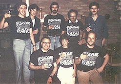 |
Note that the rhinovirus group is back row: Gert Vriend, Ming Luo, Jim Griffith, front row: Roland Rueckert, Barbara Sherry, |
Actually I had many exciting times. I mean we solved hemoglobin, lactic dehydrogenase, glyceraldehyde-3-phosphate dehydrogense. It was very exciting to see the nucleotide binding come out. But this was so wonderful and this was a functional activity on the virus. Then we saw that the escape mutations were on the surface. We didn't have many sequences of rhinoviruses as there were for poliovirus which had just been published. We saw that these were hypervariable regions and that they were on the surface and were not in the canyon. We didn't know anything about conservation of residues in the canyon but it immediately suggested why the canyon was there - namely for receptor binding.
SS So you're saying that the idea of receptor binding came immediately.
MR Yes, within days of the structure. And then we also had the idea which was later shown by Tom Smith that the antibodies - that the two arms of the antibodies would bind across the 2-fold axis and that was right.
But it must have been in early May that Eddy and Gert Vriend and I went up to Wisconsin for 2 days and we had a draft manuscript. Actually there were 2 manuscripts for Nature and we went through this together with Barbara Sherry and Roland Rueckert. We spent all of these 2 days going over it very carefully and discussing the results, getting the English right. It was a very, very nice experience and it actually was during that time that we realized, and I don't know how we realized it, that the building block of protomers which I think was first found by Baltimore and Jacobson was the VP1, VP2, VP3, VP4 unit. They were all intertwined and we saw how they were intertwined and then we saw how the pentamer units, the 12S units, were also intertwined on the 5-fold axis so we could see the assembly process happening and this happened while we were writing the paper.
When I wrote the initial draft paper, I was looking up all the foot and mouth disease virus stuff, all the polio virus stuff, Unfortunately its my habit that I don't usually read until afterwards and so I was reading all the literature and it was really a very good education for me at this point.
SS Had the receptor for rhino been identified by then?
MR No, not until 1989. So the idea that the receptor was narrow compared with an antibody was entirely interpretation, a prediction, an hypothesis, so that was ‘85. Then in ‘89 two groups Jeff Greve at Miles and independently Tim Springer's group at Harvard. That was sometime later and we got the ICAM from Jeff Greve and were doing the EM study on the complex which we are still working on.
SS And where was the paper published?.
MR Nature, but Nature took its time, very much so. They made us combine our two papers. We had one paper on the structure determination and another on the structure interpretation but they made us contract our work into one paper and they limited us in references which was very difficult as there was a lot of background. There was a lot of work that had been done and we wanted to acknowledge that our interpretation was based on other work. Actually we were greatly helped by Fred Brown in England and also by Roger Hull who I had known from the southern bean mosaic work. Without my knowing or doing anything about it they went to the Nature office and said "you have got to publish this". We actually had submitted our paper to Nature, I think, at the very end of May - May 30. It wasn't published until September.
I had given a talk somewhere- I gave a talk in England- and a talk somewhere in New York. So it was fairly well known that we solved the structure. The polio work now had a lot of help from us because Jim (Hogle) knew what to look for. It was like turnip crinkle virus which Jim worked on with Steve. He also learned how we had solved it. In his previous paper which he published not long before that, he said phase extension doesn't work. That is something which I had been working on since 1960. But he now learned from us that that was exactly how we solved it - a breakthrough in terms of technique. Then he and Dave Filman used that method and they did it extraordinarily fast. They really did a marvelous job and then together with Marie Chow wrote up the work and they submitted their paper to Science.
SS In the poliovirus work, did they have all the escape mutants? I always associate the canyon work and the escape mutants with the rhino virus
MR Oh yes, absolutely, in fact there was a meeting in Philadelphia where Barbara (Sherry) and Roland (Rueckert)were that was maybe in March of 1985 where Philip Minor was and he had escape mutations but he couldn't organize them and she basically showed him how to do it. And later, Ann Mosser from Roland's lab also helped in this, but this didn't come until much later. Jim didn't really have the advantage that we had from the marvelous work that Barbara and Roland had done on rhino. By 1985, maybe it was five years after I first talked to Roland and we had not only gone up to Wisconsin every year and the group kept getting bigger and by '85 the group was maybe 30 or 40 people.
When Philip Minor read Michael Rossmann's oral history he wrote that his "data of which there was a substantial pile had well organized long before he met Barbara Sherry at the 1985 Philadelphia meeting [see Nature (1983) 301, 674; Nature (1983) 304, 459; J. Gen. Virol. (1985), 66, 1159, and the Proceedings of the Philadelphia meeting 'Virus attachment and entry into cells', edited by R L Crowell and K Lonberg-Holm, ASM publications 1986.]"
He continued to explain, "Polio suffered from its peculiar antigenic properties. Most of the monoclonal antibodies against type 2 and type 3 are against a site which is not normally seen at all in type 1. This is hard to believe for such similar viruses and the field fell into a morass of peptides thought to be an easier way of analyzing antigenic and immunogenic sites. In fact it was seriously misleading. Thus those doing it the hard way were felt to be seriously misinterpreting their data at the time (peptides right, mutants wrong) but are now clearly thought by Michael to have seriously misinterpreted their data in an entirely different way (mass of unorganised data, etc)."
Philip Minor wrote that he thought that "the strange imbalance in immunogenicity in the virus (which is not seen to the same extent in rhinovirus) has major effects on the pathogenesis and epidemiology of polio and the type specific distribution of disease, and is therefore of absolutely no interest to x-ray crystallographers."
To read about the determination of the structure of poliovirus go to
http://virologyhistory.wustl.edu/hogle.htm
MR Another event that happened, it was in August or September of 85 was about the time that our work was actually being published that I heard from a news report that Sterling Winthrop had been doing some work on some rhino virus inhibitors. We really didn't know much about it. I got in contact with them, with Mark McKinlay and Guy Diana.
SS They were from Sterling Winthrop?
MR They came and visited us here which I remember very clearly and we were going to lunch with those two, Guy and Mark. Eddy was here and by that time Tom Smith had joined my group as a post doc. We went out for lunch to the University Inn and I remember that and they then gave us some of their inhibitors. They gave us two compounds. This was Tom's (Tom Smith) first post doc work in my lab and he diffused these into the rhino virus crystals and we took them to CHESS. It must have been the end of '85 and he collected data. Gert Vriend helped Tom process the data. He taught Tom to process the data and he then did our first maps. There were some technical problems that we solved and we saw these inhibitors in the virus. Tom first did some radioactive labeling of the compounds and showed that they actually bound to the virus and by that time we knew that they should be binding to the capsid. At first we didn't really know at what stage in the life cycle these inhibitors worked. Tom actually showed that they actually worked by binding to the capsid.
SS Did Sterling Winthrop develop these drugs?
MR I'm not quite sure of the exact story. There is a compound called arildone and there is the work from Hans Eggert (fromKoln, Germany) who had shown that similar compounds inhibited RNA synthesis and uncoating of the virus. And that's where Sterling came in.
SS Was this your first contact with the company?
MR Yes, I made the contact.
SS You had not been contacted previously by companies?
MR No, but I do remember Burroughs Wellcome contacted me wanting the coordinates which I gave them. I actually wasn't sure what to do and a number of companies wanted to buy the coordinates and I wasn't sure about the ethics. I actually wrote to Max Perutz, my former mentor, and he said: give it away and people can do with them what they like, that’s the best way. So that is what I did. By February of 1986 we had the idea with these inhibitors - and there was a meeting, a Biophysics meeting, at San Francisco when I first talked about this. Also there was a WHO meeting in Geneva which I was asked to attend where we also mentioned our work on the inhibitors. So I think that was quite significant later. It was really very informative with these inhibitors about how the virus works.
SS You mean for the first time you really understood how the inhibitors worked. And did it lead them to develop other drugs?
MR Sterling Oh, yes. You know Sterling eventually went under. It was bought by one of the French companies. They just wanted a marketing arm.
SS So those compounds are not being used?
MR No No, I got to know the scientists at Sterling very well. There was not only Guy Diana but Mark McKinlay. There was Dan Pevear and a lot of other people were involved and they then started meeting with us at the WisPur meetings. They were and are a wonderful group of people. This was my first real contact with an industrial group, but they were really good scientists and still are good scientists. And when Sterling was sold, they started a venture capital company called Viropharma, about 23 of the scientists, and continued developing these compounds. But at that time - there's a long history here - it should have happened while Sterling still existed. The first compound 51711 failed in phase I in FDA testing because it crystallized and caused damage to the urethra. The second compound - I can't even remember the name - was OK. It went through the toxicity test for phase I but it wasn't very efficacious and it was found that that compound was quickly metabolized. Subsequently it was found that the metabolites were toxic causing hepatitis. And at that time Guy Diana developed a group of about 20 synthetic organic chemists at Sterling and was synthesizing literally thousands of these compounds being guided by the structure because obviously it had to fit into the pocket.
SS At that stage were they able to do it by computers?
MR There were a lot of computer attempts but I think the best computer was Guy Diana's brain.
SS Well, you said it had to be able to fit into the pocket.
MR You could see that on the computer but it wasn't a computational drug design. There were a number of attempts at that but I think the important things were what Guy thought and said and how he developed these compounds. And his head, his brain was guided by the structure and one compound 63843 -now called pleconaril – something like that was the lead compound and about that time Sterling was sold and Viropharma existed in Philadelphia and that company is doing very well. Pleconaril has gone through phase 3 testing very successfully. It's now being used for diseases which are more serious.
SS Enteroviruses?
MR Enteroviruses: Coxsackie, echovirus and there was one particular case. I may have got this story slightly wrong but it's something like this: In Denver there was a child who was being immunized with polio vaccine but the child was immunodeficient in some way or other and the polio virus was neurovirulent and in desperation they tried this drug 63843, picornaril and the virus was cleared.
There are about 40 examples like this. They are called compassionate cases, but in fact the drug is now being used in particular for aseptic meningitis, whatever that is and for something called hand and mouth disease and various other picornavirus diseases. And now there is continual development by the company.
SS Could it be used for hepatitis A?
MR No, it probably can't be used for hepatitis A. It is not quite clear whether it can be used but there has been one guy in Sweden (Anders, I can't think of his last name) working in Lund who had worked on the use of these drugs for hepatitis A virus but it probably doesn't bind hepatitis A. It probably doesn't bind to cardioviruses like mengovirus but it does bind to the enteroviruses and to the rhinoviruses.
SS Is the structure of the cardioviruses known?
MR Yes- mengo virus we did that with Doug Scraba. You know I mentioned we collected data on that in April 1985 and in 1986 we got the structure of mengo virus.
SS So knowing the structure, would you know that this drug shouldn't fit in the pocket?
MR Yes, pretty well, but you can't be sure. One thing, when the drug goes in, the pocket actually gets bigger. It moves so you don’t know. In rhinovirus the pocket is fairly obvious but you can't be sure even if you can not see the pocket the drug can’t go in because the drug might actually cause unpredictable conformation changes.
SS So let's go back to rhinoviruses. Once you started to do rhino and had the first map did you begin to look at other picornaviruses?
MR Well we already had started doing that. In fact we were working on mengo virus. I started even before I started to work with Roland and we didn't get anywhere. I was at a meeting in Banff in Canada. I think it was on protein-protein recognition or some such subject and Doug Scraba who was in Edmonton and I happened to be sitting at a dinner table together and we talked about his work on mengo virus. One of my undergraduates actually went to his lab for a summer to try to produce mengo virus, but that didn't work out. Later Ulrike Boege, she was a graduate student in Wengler's lab, and had determined the sequence of the protein, the Sindbis capsid protein. Then she became a post doc with Doug Scraba. She had a much better idea of how to produce virus in quantity and she herself brought the virus down here and together with John Erickson took the first pictures of crystals. John left and the study evolved into a Ph.D. project for Ming Luo, one of my graduate students. Eventually he solved the structure in 1986 and then later, Jim Hogle solved the structure of Theiler's virus, that's a cardiovirus. And Ming also did another variety of Theiler's virus. He was then at the University of Alabama on the faculty. He's still there.
SS So what were some of the surprises from the rhinovirus work?
MR The biggest surprise which we didn't expect was that it was like the plant viruses, that animal viruses were like plant viruses. That was a big surprise. It wasn't immediately obvious. We saw the canyon and recognized what it was. It wasn't until about 1991 or 92 that we were able to verify that it is the receptor binding site. We were able to combine Barbara Sherry's and Roland's work with our structural work. It was a beautiful synthesis.
SS That is a very nice story.
MR And we were able to see the assembly units, the protomer.
SS But you said that didn't come until you started to write the paper
MR That's right
SS Do you remember what it was about writing the paper that allowed you to make the connections?
MR We were talking about this and probably Roland was asking about how it all fitted together in 3 dimensions. In the map you only see one asymmetric unit but it’s the relationship between the asymmetric units here that is important. I remember suddenly it came to all of us as we were talking.
| Introduction
| Some historical highlights: structural virology
and virology |
| Solving the Structure of Icosahedral Plant Viruses
| Picornavirus Structure | Poliovirus
| Polio
The Influenza Virus Hemagglutinin | The
Influenza Virus Neuraminidase
| Issues of Science and Society |
contributors| Home |